44 labeled cell diagram
anatomy cell structure Prostate Structure And Function | Abdominal Key abdominalkey.com. prostate structure function abdominal. Pin On Monirqa . cell animal functions structure diagram labeled parts plant shape grade function 6th organelles membrane buzzle. Structure Of The Generalized Cell Quiz . cell structure quizlet quiz ... 140,336 Labelled cell Images, Stock Photos & Vectors - Shutterstock 140,275 labelled cell stock photos, vectors, and illustrations are available royalty-free. See labelled cell stock video clips Image type Orientation People Artists Sort by Popular Biology Healthcare and Medical Science Animals and Wildlife cell anatomy eukaryote molecule organelle nucleus Next of 1,403
cell diagram to label Label The Cell Diagram . cell golgi membrane label diagram animal bodies apparatus plasma nucleus 6th facts cells organelles inside ribosomes project which structures genetic. 34 Human Cell Diagram To Label - Labels For Your Ideas opilizeb.blogspot.com. quizlet. A Typical Cell, Labeled Stock Photo 112395266 : Shutterstock

Labeled cell diagram
CELL MEMBRANE LABEL Diagram | Quizlet Practice labeling the parts of the cell membrane Terms in this set (6) Channel Protein hole or tunnel that particles may pass through to go in / out of cell Marker protein identifies or labels the cell Receptor protein receives information Heads part of the phospholipid that loves water (hydrophili) - points to the most outside and inside of cell Cell Structure | SEER Training Cell Structure. Ideas about cell structure have changed considerably over the years. Early biologists saw cells as simple membranous sacs containing fluid and a few floating particles. Today's biologists know that cells are infinitely more complex than this. There are many different types, sizes, and shapes of cells in the body. Cell: Structure and Functions (With Diagram) 1. Eukaryotes are sophisticated cells with a well defined nucleus and cell organelles. 2. The cells are comparatively larger in size (10-100 μm). 3. Unicellular to multicellular in nature and evolved ~1 billion years ago. 4. The cell membrane is semipermeable and flexible. 5.
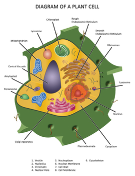
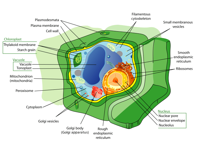
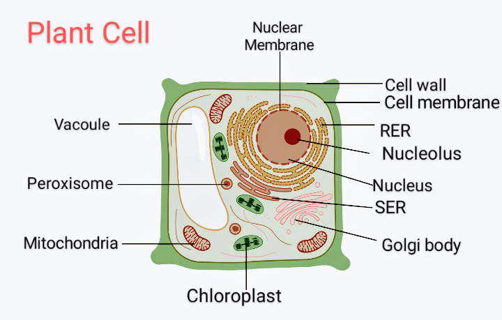
Labeled cell diagram. A Labeled Diagram of the Plant Cell and Functions of its Organelles A Labeled Diagram of the Plant Cell and Functions of its Organelles We are aware that all life stems from a single cell, and that the cell is the most basic unit of all living organisms. The cell being the smallest unit of life, is akin to a tiny room which houses several organs. Here, let's study the plant cell in detail... Cell Diagrams - The Biology Corner This will add text boxes that can be filled in. You can also use drop-down fields if you want to provide students with choices in how to label the diagram. Students will need to download the pdf in order to fill in the fields. Open Google Draw and import the diagram. Then use "insert" to create text boxes where students can fill in the labels. Human Cell Diagram, Parts, Pictures, Structure and Functions One of the few cells in the human body that lacks almost all organelles are the red blood cells. The main organelles are as follows : cell membrane endoplasmic reticulum Golgi apparatus lysosomes mitochondria nucleus perioxisomes microfilaments and microtubules Diagram of the human cell illustrating the different parts of the cell. Cell Membrane Label the cell - Teaching resources - Wordwall Label Plant and Animal Cell Labelled diagram by Eawilson the cell Match up by Elenagp9149 5.6 Label the sentence Labelled diagram by Christianjolene Label the Electromagnetic Spectrum Labelled diagram by Elizabetheck G6 G7 G8 Science 5.7 Label the sentence Labelled diagram by Christianjolene The cell Anagram by Thepowerhouse G7 G8 Science
liver diagram labeled pig fetal digestive dissection system anatomy external biology. 2.3.1 Draw And Label A Diagram Of The Ultrastructure Of A Liver Cell As . liver cell drawing diagram draw label ultrastructure paintingvalley animal example labelling diagrams ze kig source. This Diagrams Shows The Major Arteries In The Human Body Dieses www ... Cell Cycle Diagram Blank - amy brown science teaching cellular ... Cell Cycle Diagram Blank - 17 images - fill in the blank water cycle diagram worksheet, the cell cycle diagram labeled how to visualize data in your, biology for grade 11, academic biology, ... Cell Membrane Unlabeled Labeled Cell Diagram, Media.nbcmontana.com is an open platform for users to share their favorite wallpapers, By downloading this ... prokaryotic cell diagram worksheet cell labeling science worksheets eukaryotic activities plant prokaryotic cells structure graders 1st bing. Cell structure function cells prokaryotic microbiology label focus labels worksheet related typical approach looking. Bacteria structure bacterial notes biologycorner. Bacterial cell Structure of Cell: Definition, Types, Diagram, Functions - Embibe Structure of Cell: Cell is the basic functional unit that makes up all living organisms.All organisms, including ourselves, start life as a single cell called the egg. Cells are small microscopic units that perform all essential functions of life and are capable of independent existence.
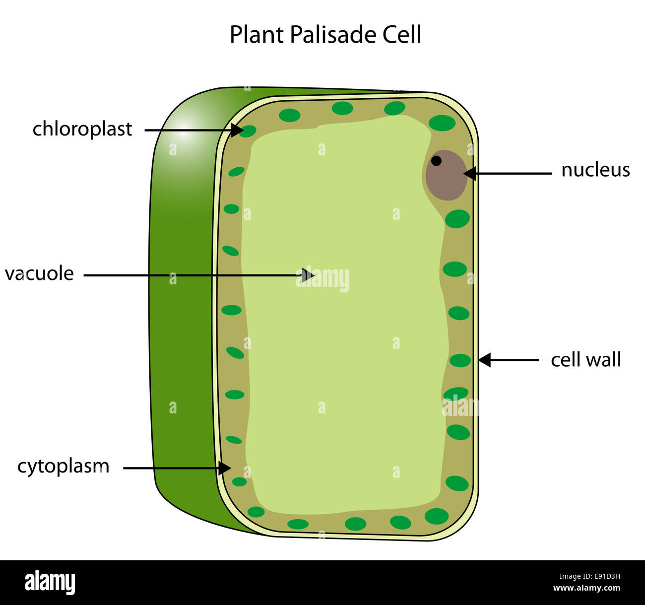
What Is Going On Inside That Cell? | Human cell diagram, Cell diagram ... Cells are the basic building blocks of living things. The human body is composed of trillions of cells, all with their own specialised function. The animal cell diagram. Vector illustration on white background. Here is a big collection of biology worksheets for you to use with your students. PDF Human Cell Diagram, Parts, Pictures, Structure and Functions Diagram of the human cell illustrating the different parts of the cell. Cell Membrane The cell membraneis the outer coating of the cell and contains the cytoplasm, substances within it and the organelle. It is a double-layered membrane composed of proteins and lipids. Learn the parts of a cell with diagrams and cell quizzes Cell diagram unlabeled It's time to label the cell yourself! As you fill in the cell structure worksheet, remember the functions of each part of the cell that you learned in the video. Doing this will help you to remember where each part is located. Click the links below to download the labeled and unlabeled eukaryotic cell diagrams. Labeled Plant Cell With Diagrams | Science Trends The parts of a plant cell include the cell wall, the cell membrane, the cytoskeleton or cytoplasm, the nucleus, the Golgi body, the mitochondria, the peroxisome's, the vacuoles, ribosomes, and the endoplasmic reticulum. Parts Of A Plant Cell The Cell Wall Let's start from the outside and work our way inwards.
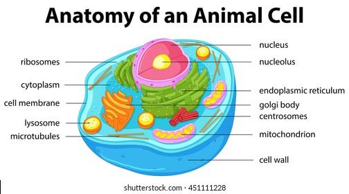
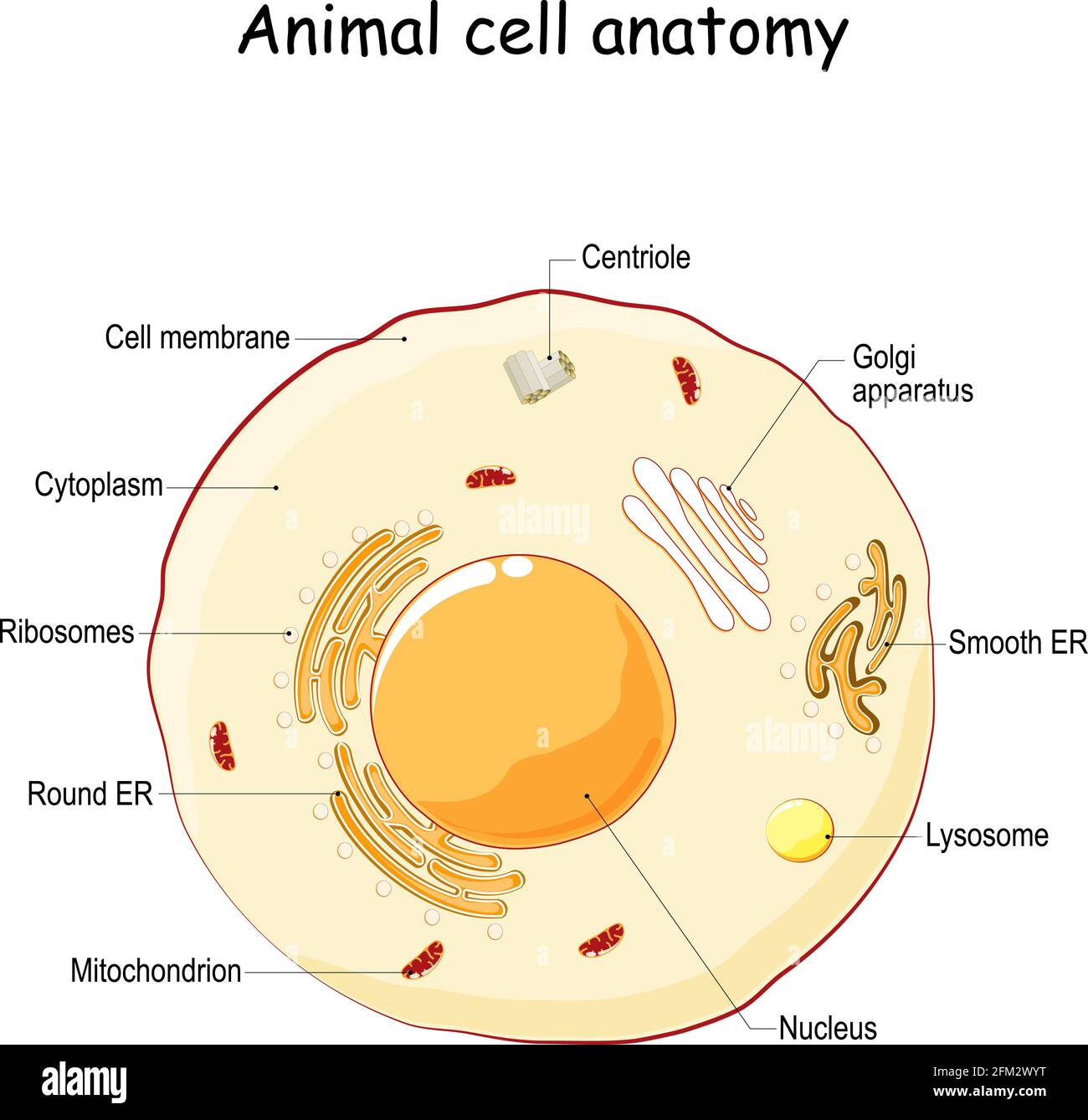
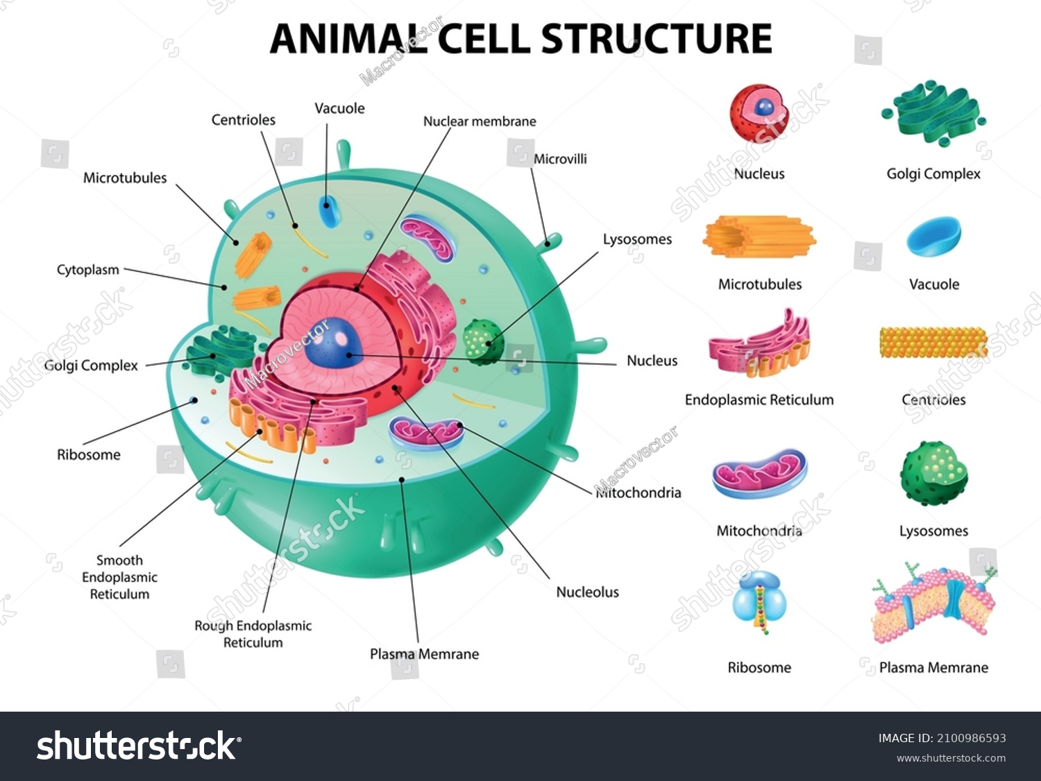
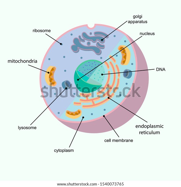
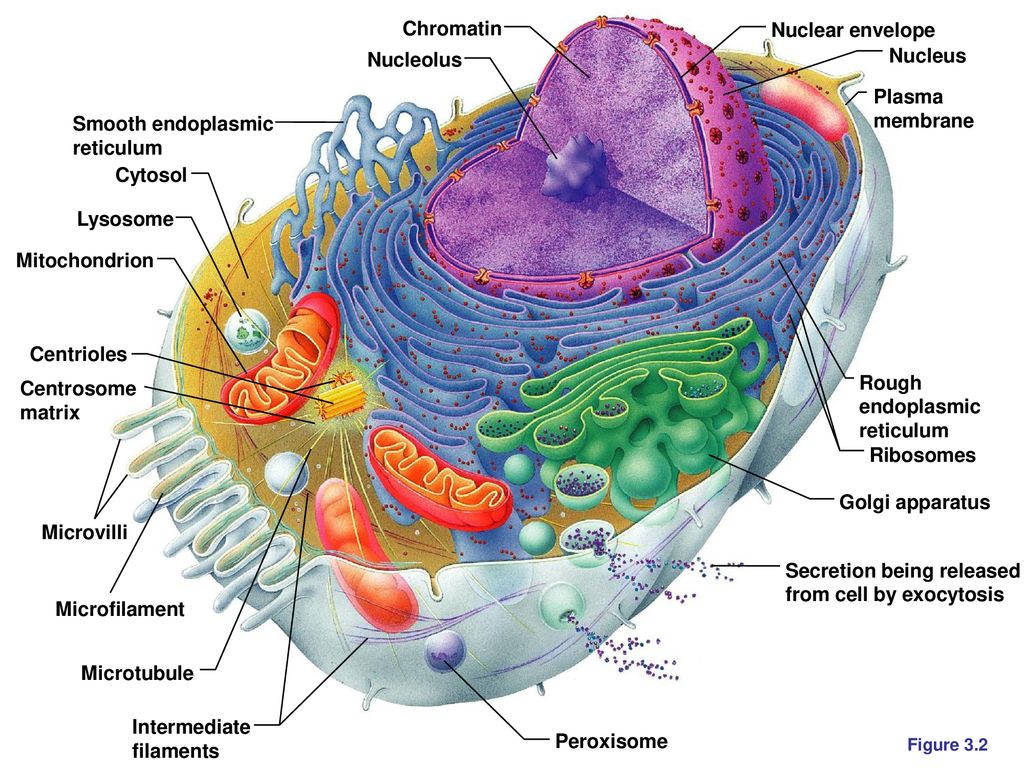
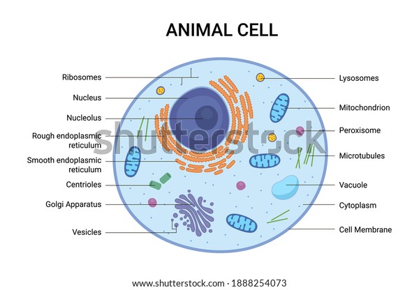
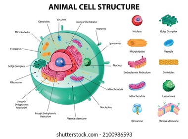
A Well-labelled Diagram Of Animal Cell With Explanation Well-Labelled Diagram of Animal Cell The Cell Organelles are membrane-bound, present within the cells. There are various organelles present within the cell and are classified into three categories based on the presence or absence of membrane. Listed below are the Cell Organelles of an animal cell along with their functions.
Bacteria in Microbiology - shapes, structure and diagram The bacteria shapes, structure, and labeled diagrams are discussed below. Sizes The sizes of bacteria cells that can infect human beings range from 0.1 to 10 micrometers. Some larger types of bacteria such as the rickettsias, mycoplasmas, and chlamydias have similar sizes as the largest types of viruses, the poxviruses.
Plant Cell: Meaning, Components, Structure, Functions & Parts - Embibe It is a rigid layer that is composed of cellulose, glycoproteins, lignin, pectin and hemicellulose. It is located outside the cell membrane and is completely permeable. The primary function of a plant cell wall is to protect the cell against mechanical stress and to provide a definite form and structure to the cell.
A Labeled Diagram of the Animal Cell and its Organelles A Labeled Diagram of the Animal Cell and its Organelles There are two types of cells - Prokaryotic and Eucaryotic. Eukaryotic cells are larger, more complex, and have evolved more recently than prokaryotes. Where, prokaryotes are just bacteria and archaea, eukaryotes are literally everything else.
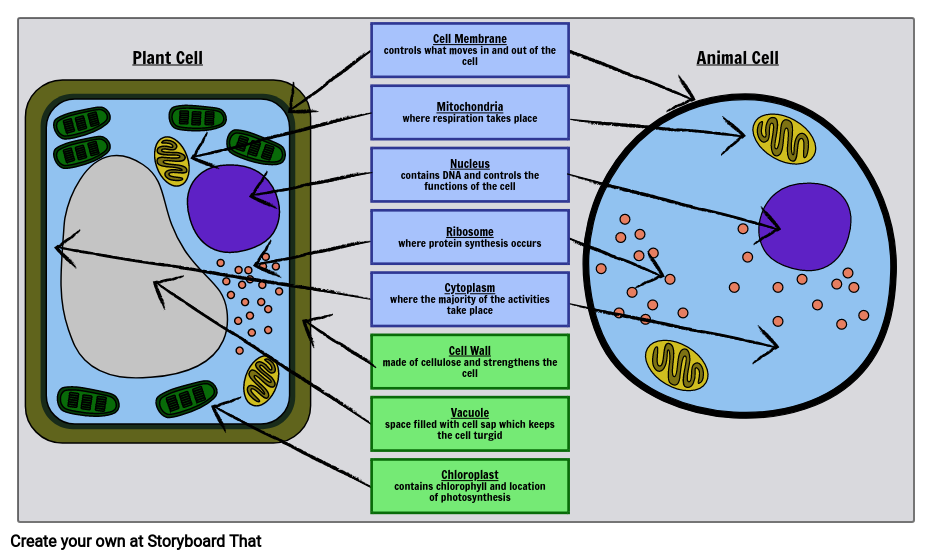
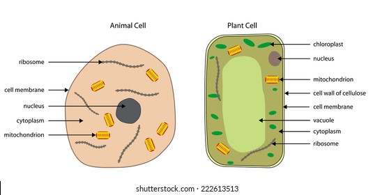
Animal Cells: Labelled Diagram, Definitions, and Structure Plant Cells Diagram Plant Cells Organelles and Functions Animals Cells Vs Plant Cells Plant and animal cells have several differences such as plant cells have a cell wall or chloroplasts, but animal cells do not have either. Plant cells are fixed, rectangular in shapes, but animal cells are mostly round and irregular in shape.
Animal Cell Diagram with Label and Explanation: Cell Structure, Functions Diagram of Animal Cell Below is the diagram of the animal cell which shows the organelles present in it. The cell is covered with cytoplasm which consists of cell organelles in it. The nucleus is covered with a rough Endoplasmic Reticulum and other organelles each designed for a specific purpose.
03 Label the Cell Diagram | Quizlet Start studying 03 Label the Cell. Learn vocabulary, terms, and more with flashcards, games, and other study tools.
Cells Diagram | Science Illustration Solutions - Edrawsoft Cells Diagram. Cells are the basic building blocks of all living things. The human body is composed of trillions of cells. Cells have many parts, each with a different function. Some of these parts, called organelles, are specialized structures that perform certain tasks within the cell. Drawing cells diagram helps you better understand your ...
Cell Organelles- Definition, Structure, Functions, Diagram An additional non-living layer present outside the cell membrane in some cells that provides structure, protection, and filtering mechanism to the cell is the cell wall. Structure of Cell Wall. In a plant cell, the cell wall is made up of cellulose, hemicellulose, and proteins while in a fungal cell, it is composed of chitin. A cell wall is ...
Structure of Bacterial Cell (With Diagram) - Biology Discussion It is 10-25 nm in thickness. It gives shape to the cell. Nucleus: The single circular double-stranded chromosome is the bacterial genome. Other structures include cytoplasmic membrane, mesosomes, ribosomes and cytoplasmic inclusions. Unlike eukaryotes cytoplasm does not contain ribosome, Golgi, cytoskeleton.
Cell: Structure and Functions (With Diagram) 1. Eukaryotes are sophisticated cells with a well defined nucleus and cell organelles. 2. The cells are comparatively larger in size (10-100 μm). 3. Unicellular to multicellular in nature and evolved ~1 billion years ago. 4. The cell membrane is semipermeable and flexible. 5.
Cell Structure | SEER Training Cell Structure. Ideas about cell structure have changed considerably over the years. Early biologists saw cells as simple membranous sacs containing fluid and a few floating particles. Today's biologists know that cells are infinitely more complex than this. There are many different types, sizes, and shapes of cells in the body.
CELL MEMBRANE LABEL Diagram | Quizlet Practice labeling the parts of the cell membrane Terms in this set (6) Channel Protein hole or tunnel that particles may pass through to go in / out of cell Marker protein identifies or labels the cell Receptor protein receives information Heads part of the phospholipid that loves water (hydrophili) - points to the most outside and inside of cell

(434).jpg)














Post a Comment for "44 labeled cell diagram"