38 light microscope diagram with labels
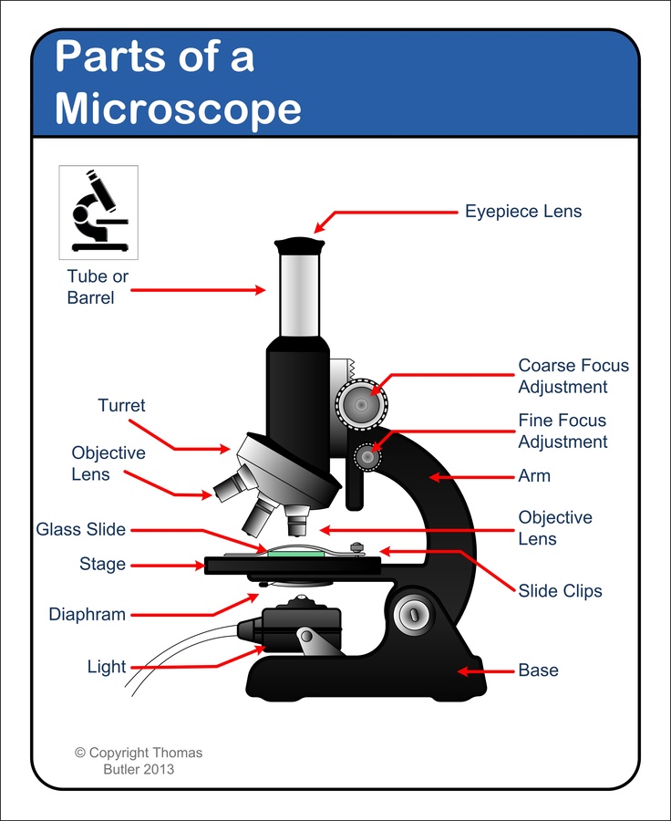
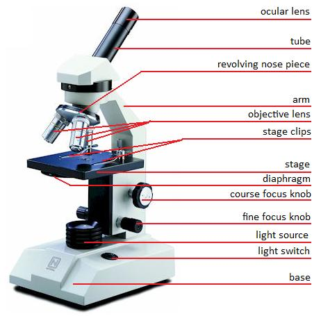
Label Microscope Diagram - EnchantedLearning.com low-power objective - a small lens with low magnifying power. mirror (or light source) - this directs light upwards onto the slide. revolving nosepiece - the rotating device that holds the objectives (lenses). stage - the platform on which a slide is placed. stage clips - metal clips that hold a slide securely onto the stage. Label the Light Microscope - Labelled diagram - Wordwall Label the Light Microscope - Labelled diagram Eyepiece, Light Source, Base, Stage, Stage Clips, Fine Focus, Coarse Focus, Arm, Objective Lens. Label the Light Microscope Share by Nquinn805 Like Edit Content More Leaderboard Switch template
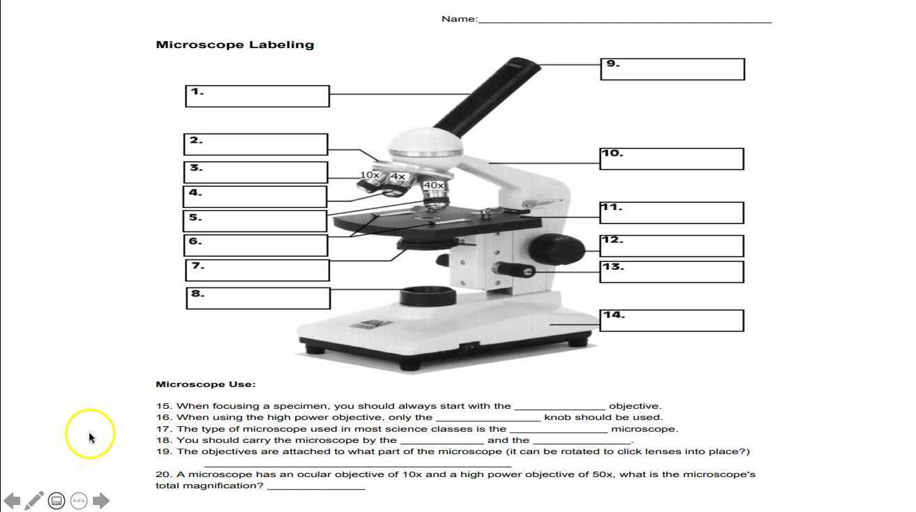
Light Microscope: Functions, Parts and How to Use It To use a light microscope, you can follow the steps below carefully. Start with a low lens and a clean slide. The microscope stage should be lowered as low as possible. Center the slide so that the specimen is under the objective lens. Use the coarse adjustment knob to get a general focus. Then slowly move up the stage until focus is achieved.

Light microscope diagram with labels
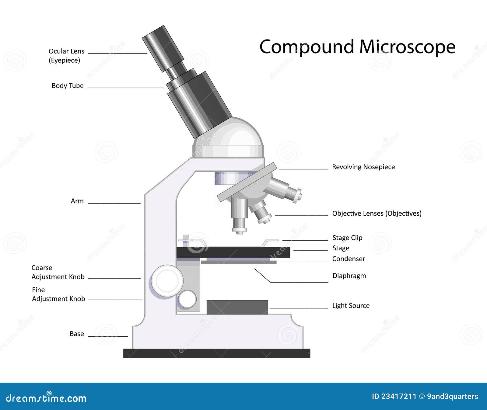
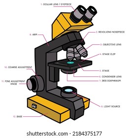
Compound Microscope Parts, Functions, and Labeled Diagram Compound Microscope Definitions for Labels. Eyepiece (ocular lens) with or without Pointer: The part that is looked through at the top of the compound microscope. Eyepieces typically have a magnification between 5x & 30x. Monocular or Binocular Head: Structural support that holds & connects the eyepieces to the objective lenses. A Study of the Microscope and its Functions With a Labeled Diagram ... These labeled microscope diagrams and the functions of its various parts, attempt to simplify the microscope for you. However, as the saying goes, 'practice makes perfect', here is a blank compound microscope diagram and blank electron microscope diagram to label. Download the diagrams and practice labeling the different parts of these ... 16 Parts of a Compound Microscope: Diagrams and Video The eyepiece, also known as the "Ocular", is the first magnification lens you will look through in a compound microscope. Put simply, this is where you put your eye to see the image. Usually eyepieces come in 10X magnification, or 15X magnification but they can vary from 5X - 30X.
Light microscope diagram with labels. Labeling the Parts of the Microscope | Microscope World Resources Labeling the Parts of the Microscope This activity has been designed for use in homes and schools. Each microscope layout (both blank and the version with answers) are available as PDF downloads. You can view a more in-depth review of each part of the microscope here. Download the Label the Parts of the Microscope PDF printable version here. Compound Microscope Parts - Labeled Diagram and their Functions The eyepiece (or ocular lens) is the lens part at the top of a microscope that the viewer looks through. The standard eyepiece has a magnification of 10x. You may exchange with an optional eyepiece ranging from 5x - 30x. [In this figure] The structure inside an eyepiece. The current design of the eyepiece is no longer a single convex lens. Labelled Diagram Of A Light Microscope | Products & Suppliers ... A schematic diagram for the microscope -based label -free microfluidic light scattering cytometer. IB Biology/Option H - Further Human Physiology - Wikibooks, open books for an open world Draw and label a diagram showing a transverse section of the ileum as seen under a light microscope . Sperm Under Microscope with Labeled Diagram - AnatomyLearner All these structures are identified in the seminiferous tubules 400x labeled diagram. Okay, let's see the main difference between the different types of spermatogenic cells and sperm. Spermatogonia under the light microscope In the germinal epithelium of a seminiferous tubule, you will find spermatogonia (stem cells) at its base.
Light microscopes - Cell structure - Edexcel - BBC Bitesize The components of a light microscope and their functions Calculating the magnification of light microscopes. The compound microscope uses two lenses to magnify the specimen: the eyepiece and an ... Microscope, Microscope Parts, Labeled Diagram, and Functions The Iris Diaphragm is located above the condenser lens and below the microscope stage. The different sized holes in the diaphragm helps to vary the size of the cone and intensity of light that is projected upward into the slide. However, there is no set rule regarding which setting to use for a particular power. Parts of the Microscope with Labeling (also Free Printouts) Parts of the Microscope with Labeling (also Free Printouts) By Editorial Team March 7, 2022 A microscope is one of the invaluable tools in the laboratory setting. It is used to observe things that cannot be seen by the naked eye. Table of Contents 1. Eyepiece 2. Body tube/Head 3. Turret/Nose piece 4. Objective lenses 5. Knobs (fine and coarse) 6. Label the microscope Diagram | Quizlet Diaphragm. Regulates the amount of light on the specimen. Light Source. Projects light upwards through the diaphragm, the specimen, and the lenses. Arm. supports the body tube. Stage. Supports the slide being viewed. Coarse Adjustment.
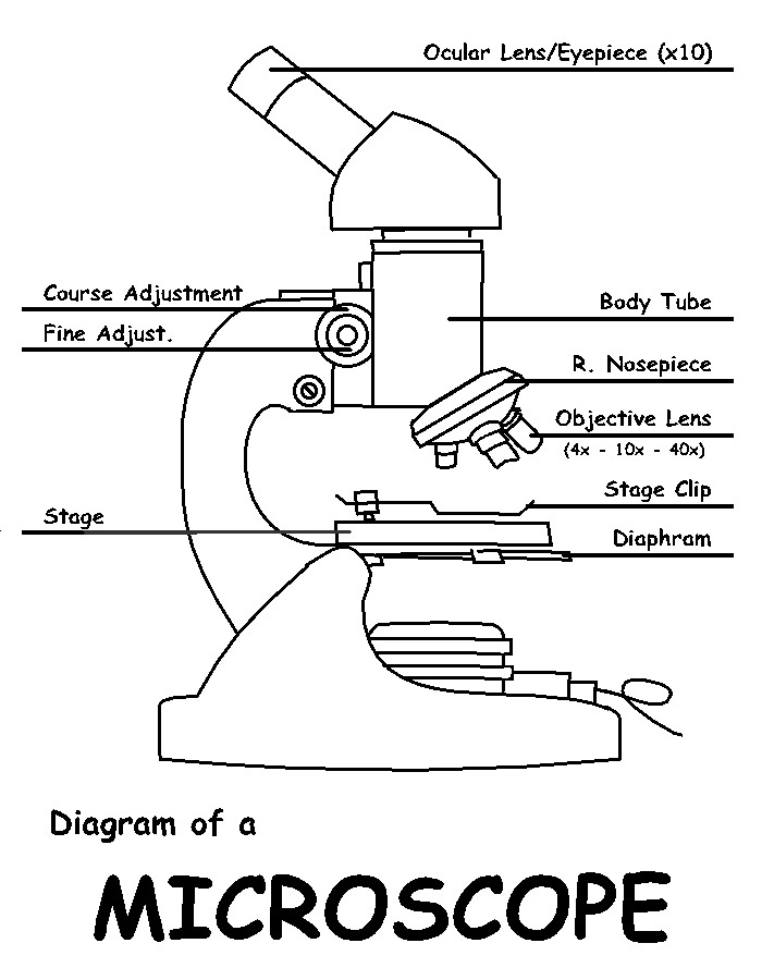
Microscope Parts, Function, & Labeled Diagram - slidingmotion Condenser. The condenser is to focus the light, which passes from the microscopic illuminator to the specimen. This condenser is located just below the diaphragm. This diaphragm is one of the important parts of the compound microscope which will help to get an accurate and sharp image. The condenser has a magnification power of 400X and above. Parts of a microscope with functions and labeled diagram - Microbe Notes Figure: Diagram of parts of a microscope There are three structural parts of the microscope i.e. head, base, and arm. Head - This is also known as the body. It carries the optical parts in the upper part of the microscope. Base - It acts as microscopes support. It also carries microscopic illuminators. Labeled Microscope and Basics of Life Diagram | Quizlet PLAY. A microscope is an instrument widely to magnify and resolve the image of an object that is otherwise invisible to naked eye. For resolving the details of objects, which otherwise cannot be achieved by naked eye, a microscope is used. This set of flash cards will help the student to identify the different parts and function of the microscope. PDF Parts of the Light Microscope - Science Spot Supports the MICROSCOPE D. STAGE CLIPS HOLD the slide in place C. OBJECTIVE LENSES Magnification ranges from 10 X to 40 X F. LIGHT SOURCE Projects light UPWARDS through the diaphragm, the SPECIMEN, and the LENSES H. DIAPHRAGM Regulates the amount of LIGHT on the specimen E. STAGE Supports the SLIDE being viewed K. ARM Used to SUPPORT the
Labeling the Parts of the Microscope | Microscope World Resources
Label the microscope — Science Learning Hub Use this with the Microscope parts activity to help students identify and label the main parts of a microscope and then describe their functions. Drag and drop the text labels onto the microscope diagram. If you want to redo an answer, click on the box and the answer will go back to the top so you can move it to another box.

Parts of a Microscope | Back to Microscopy | Biology notes ...
Microscope Labeling Game - PurposeGames.com About this Quiz. This is an online quiz called Microscope Labeling Game. There is a printable worksheet available for download here so you can take the quiz with pen and paper. This quiz has tags. Click on the tags below to find other quizzes on the same subject. Science.

GCSE Optical microscope labelling Diagram | Quizlet
Microscope Types (with labeled diagrams) and Functions Simple microscope labeled diagram Simple microscope functions It is used in industrial applications like: Watchmakers to assemble watches Cloth industry to count the number of threads or fibers in a cloth Jewelers to examine the finer parts of jewelry Miniature artists to examine and build their work Also used to inspect finer details on products

Parts of a microscope with functions and labeled diagram
Labelled Diagram of Compound Microscope The below mentioned article provides a labelled diagram of compound microscope. Part # 1. The Stand: The stand is made up of a heavy foot which carries a curved inclinable limb or arm bearing the body tube. The foot is generally horse shoe-shaped structure (Fig. 2) which rests on table top or any other surface on which the microscope in kept.
Free Microscope Drawing, Download Free Microscope Drawing png ...
Parts of Stereo Microscope (Dissecting microscope) - labeled diagram ... Labeled part diagram of a stereo microscope Major structural parts of a stereo microscope There are three major structural parts of a stereo microscope. The viewing Head includes the upper part of the microscope, which houses the most critical optical components, including the eyepiece, objective lens, and light source of the microscope.

Microscope Maintenance Tips | Science supplies, Microscope ...
Microscope Labeling - The Biology Corner 1) Start with scanning (the shortest objective) and only use the COARSE knob . Once it is focused… 2) Switch to low power (medium) and only use the COARSE knob . You may need to recenter your slide. Once it is focused.. 3) Switch to high power (long objective).

Microscope With Labels Clip Art at Clker.com - vector clip ...
Microscope Parts and Functions First, the purpose of a microscope is to magnify a small object or to magnify the fine details of a larger object in order to examine minute specimens that cannot be seen by the naked eye. Here are the important compound microscope parts... Eyepiece: The lens the viewer looks through to see the specimen.

22 Parts Of a Microscope With Their Function And Labeled ...
Light Microscope- Definition, Principle, Types, Parts, Labeled Diagram ... A light microscope is a biology laboratory instrument or tool, that uses visible light to detect and magnify very small objects and enlarge them. They use lenses to focus light on the specimen, magnifying it thus producing an image. The specimen is normally placed close to the microscopic lens.
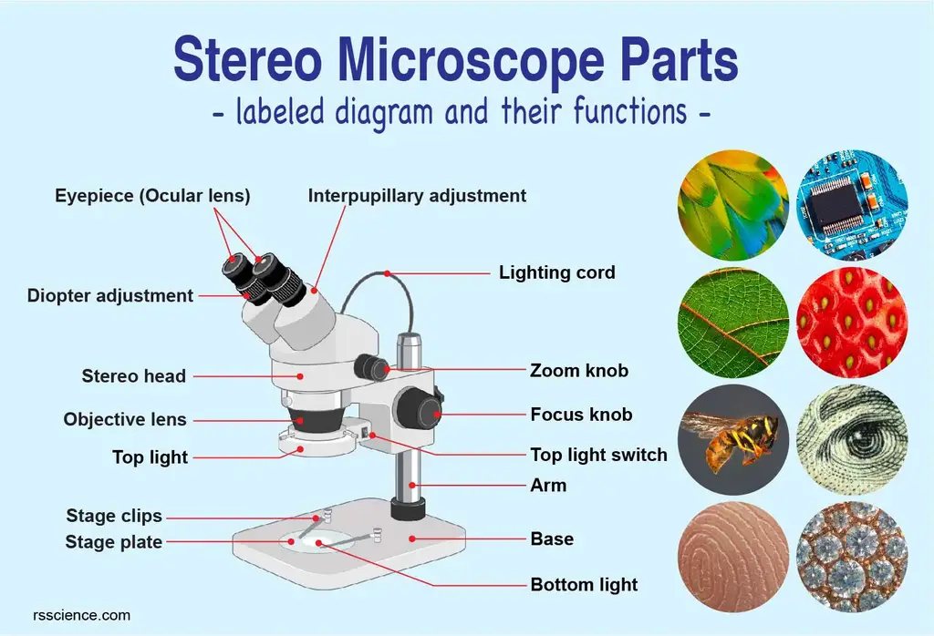
Parts of Stereo Microscope (Dissecting microscope) – labeled ...
Simple Squamous Epithelium under a Microscope with a Labeled Diagram ... I think you got the basic idea of the simple squamous epithelium under the light microscope. So, the simple squamous epithelium consists of a single layer of spindle-shaped (flattened) cells, a tapered or elongated nucleus that rests on a basement membrane. All the labeled diagrams of simple squamous epithelium might help you learn them.
Label a microscope - Teaching resources
Label Microscope Diagram - EnchantedLearning.com Using the terms listed below, label the microscope diagram. arm - this attaches the eyepiece and body tube to the base. base - this supports the microscope. body tube - the tube that supports the eyepiece. coarse focus adjustment - a knob that makes large adjustments to the focus. diaphragm - an adjustable opening under the stage, allowing ...

Biology Microscope Labeling and Definitions (Light/Compound ...
Simple Microscope - Diagram (Parts labelled), Principle, Formula and Uses Parts of a Simple Microscope A simple microscope consists of Optical parts Mechanical parts Labeled Diagram of simple microscope parts Optical parts The optical parts of a simple microscope include Lens Mirror Eyepiece Lens A simple microscope uses biconvex lens to magnify the image of a specimen under focus.
Microscope, Microscope Parts, Labeled Diagram, and Functions
16 Parts of a Compound Microscope: Diagrams and Video The eyepiece, also known as the "Ocular", is the first magnification lens you will look through in a compound microscope. Put simply, this is where you put your eye to see the image. Usually eyepieces come in 10X magnification, or 15X magnification but they can vary from 5X - 30X.

Solved A. OLYMPUS C. B. Use the Diagram to answer the | Chegg.com
A Study of the Microscope and its Functions With a Labeled Diagram ... These labeled microscope diagrams and the functions of its various parts, attempt to simplify the microscope for you. However, as the saying goes, 'practice makes perfect', here is a blank compound microscope diagram and blank electron microscope diagram to label. Download the diagrams and practice labeling the different parts of these ...

Compound Microscope Parts, Diagram Definition, Application ...
Compound Microscope Parts, Functions, and Labeled Diagram Compound Microscope Definitions for Labels. Eyepiece (ocular lens) with or without Pointer: The part that is looked through at the top of the compound microscope. Eyepieces typically have a magnification between 5x & 30x. Monocular or Binocular Head: Structural support that holds & connects the eyepieces to the objective lenses.

Label the microscope — Science Learning Hub

Leica Upright Compound Light Microscope Diagram Diagram | Quizlet

Schematic diagrams of (a) in-situ optical measurement system ...

Label The Microscope Diagram - Robot PNG Image | Transparent ...
Parts of a Microscope with Their Functions • Microbe Online

Compound Microscope Parts – Labeled Diagram and their ...
Label the Light Microscope - Diagram berlabel

MICROSCOPE Labeling - Part - 3

Labels for the light microscope for... - The Science Break ...
Compound Microscope Stock Illustrations – 726 Compound ...
biology labeled microscope diagram - Clip Art Library
Microscope Labeling

easy compound microscope diagram - Clip Art Library
The Microscope

Compound microscope labeling Diagram | Quizlet

Microscope With Labels clip art | Microscope parts ...
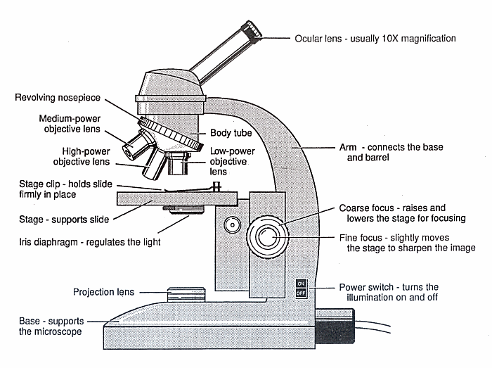
Diagram of a Microscope - Guide to using a microscope

Microscope Diagram Labeled, Unlabeled and Blank | Parts of a ...

monocular labeling Diagram | Quizlet
Compound Light Microscope Labeled - ClipArt Best

Microscope labeled diagram

Simple Microscope - Diagram (Parts labelled), Principle ...
10,528 Light microscopy Images, Stock Photos & Vectors ...

label microscope diagram | Charts | Microscope, Anatomy bones ...
Post a Comment for "38 light microscope diagram with labels"