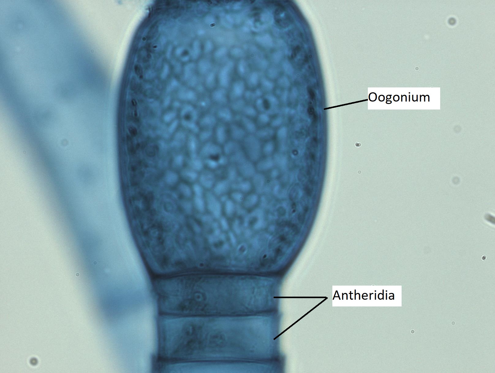
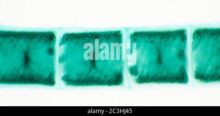
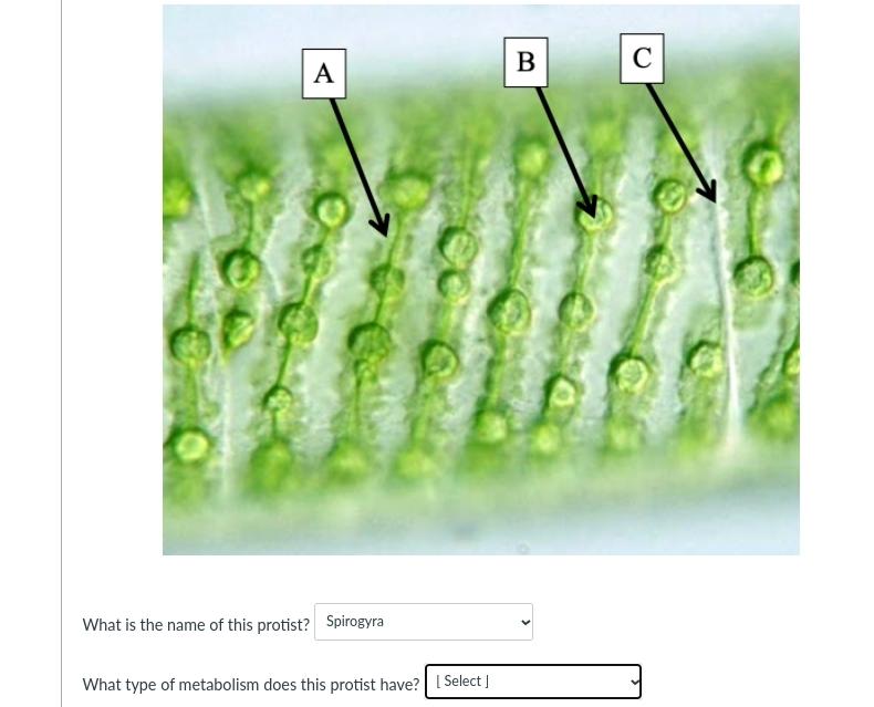
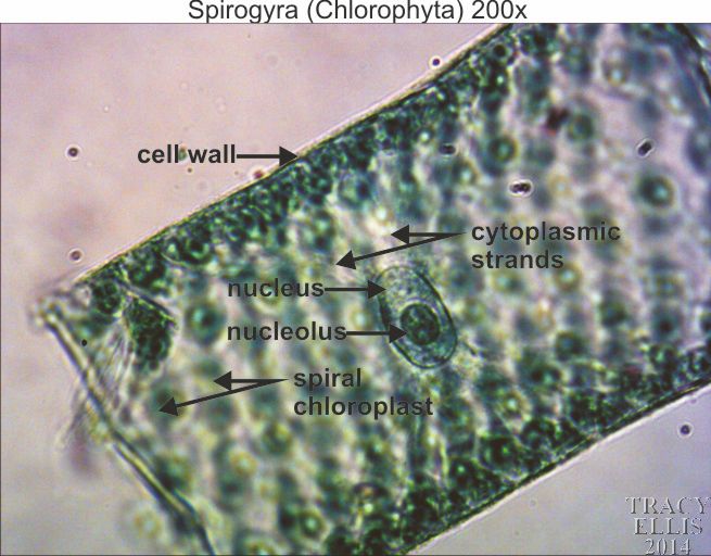
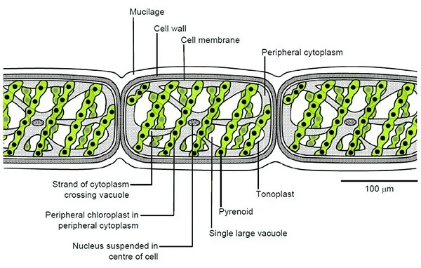
38 spirogyra microscope labeled
Spirogyra Flashcards | Quizlet common name for spirogyra due to its slippery touch. structure. multicellular, long filamentous body composed of many similar cells. cell wall. outermost covering made of cellulose with pectin on the outerside. pectin. gets sticky in water, causes spirogyra to be slippery. cell membrane. lies under cell wall and is made of protein and lipid ... Spirogyra, vegetative, multiple chloroplasts, WM Microscope slide ... Prepared microscope slide of Spirogyra, vegetative, multiple chloroplasts, WM
Algae lab - The Biology Corner Sketch the spirogyra. LABEL the chloroplasts and the cell wall. 10. View the volvox under scanning and low power. Sketch the volvox. Post-Lab Analysis 11. Compare and Contrast each of the 3 Algae you looked at by filling out a Venn Diagram (you should be able to come up with at least 3 items for each group)

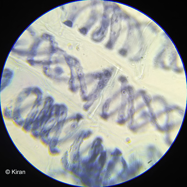
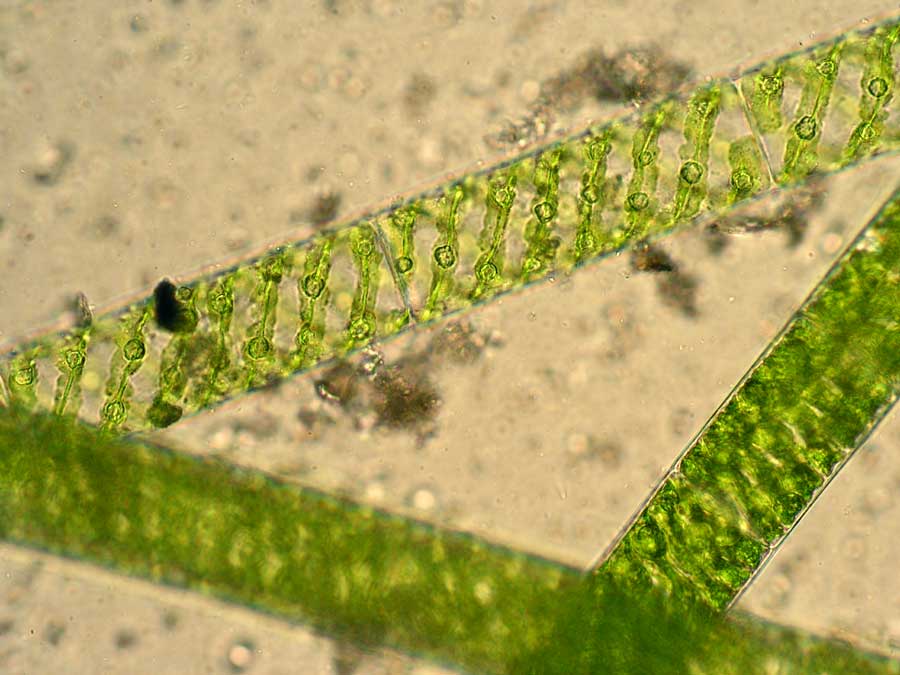
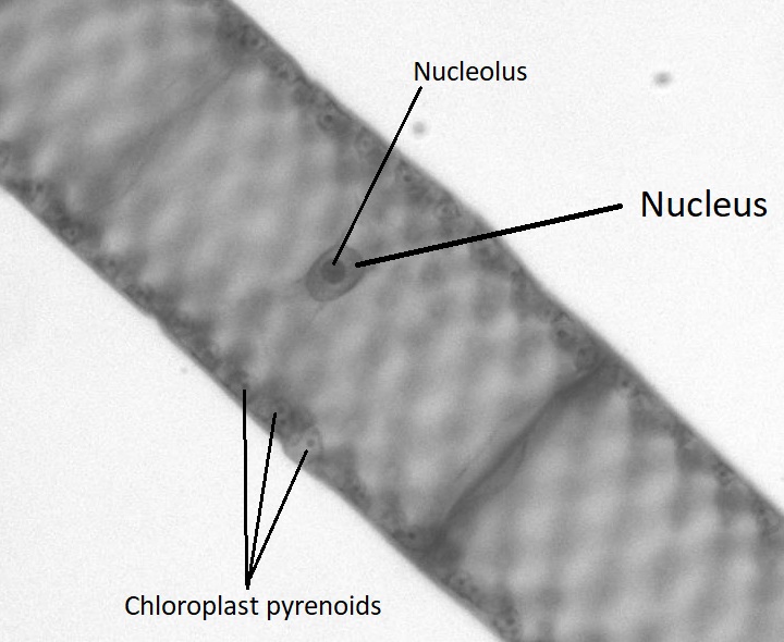
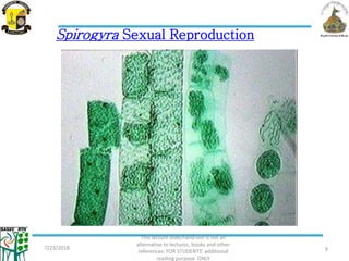
Spirogyra microscope labeled
Spirogyra - Under the Microscope - YouTube Music: C Major Prelude - Bach 1.5: Microscopy - Biology LibreTexts Review the following rules and tips for using and handling your microscope. Figure 1. Labeled parts of a microscope. General Rules Always START and END with the low power lens when putting on OR taking away a slide. Never turn the nose piece by the objective lens. Biological drawing. Spirogyra Filament. Resources for Biology Education ... Spirogyra Filament. Spirogya is a filamentous alga. Its cells form long, thin strands that, in vast numbers, contribute to the familiar green, slimy 'blanket weed' in ponds. Seen under the microscope, each filament consists of an extensive chain of identical cells.
Spirogyra microscope labeled. Microscope Imaging Station. Gallery. - Exploratorium Spirogyra, structures labeled This algae grows in long, hairlike strands in freshwater ponds. The helical green structures inside the cell walls are chloroplasts. More images in this category: Elodea leaf cells with structures labeled Spirogyra, structures labeled Sea urchin embryo cell division, organelles labeled Genus Spirogyra - An Overview - Microbe Notes Under the microscope, Spirogyra appears surrounded by a slimy jelly-like substance which is the outer wall of the organism dissolved in water. Besides, the filaments are also surrounded by mucilage that holds the filaments together to form clumps in water. ... Functions, Labeled Diagram; Normal Flora (Microbiota) of Mouth and Gastrointestinal ... Spirogyra Labelled Diagram Spirogyra (common names include water silk, mermaid's tresses, and blanket weed) is a genus of filamentous charophyte green algae of the order Zygnematales, named for the helical or spiral arrangement of the chloroplasts that is characteristic of the genus. Draw a labelled diagram of Spirogyra. 51 Differentiate between flying lizard and bird. Freshwater Algae: Filamentous: Page 1. Introductory text with ... Spirogyra, Oedogonium and Cladophora are amongst the varieties most frequently encountered. All blue-green algae are now classified amongst the Bacteria, and will be found in the Cyanobacteria gallery. Spirogyra. Spirogyra is a filamentous green alga which is common in freshwater habitats. It has the appearance of very fine bright dark-green ...
Amazing 27 Things Under The Microscope With Diagrams May 13, 2022 · 23. Spirogyra under the microscope. Spirogyra is a green alga found mostly in freshwater in the form of green clumps. Spirogyra is unicellular, but because it clumps together, it can be seen in the pond even with our naked eyes. These organisms have green pigments that are arranged in the form of ribbons in the cytoplasm. Spirogyra - Wikipedia Spirogyra (common names include water silk, mermaid's tresses, and blanket weed) is a filamentous charophyte green alga of the order Zygnematales, named for the helical or spiral arrangement of the chloroplasts that is characteristic of the genus. Spirogyra Prepared Microscope Slide - Acorn Naturalists Spirogyra (prepared microscope slide) Share how you use this resource $4.95 Item T-15178 Qty Add to Cart Add to Wish List Add to Compare Email Print this Page Spirogyra Prepared Microscope Slide Named for the helical or spiral arrangement of the chloroplasts. It is commonly found in freshwater habitats where it may form large floating mats. Spirogyra Microscope Slides, w.m. | Carolina.com Spirogyra Single Chloroplast, w.m. Microscope Slide #296554 (in stock) $5.45 Quantity Discount Available Quantity add to wishlist Spirogyra Scalariform Conjugation, Vegetative and Zygote Stages, w.m. Microscope Slide #296560 (in stock) $7.60 Quantity Discount Available Quantity add to wishlist
Spirogyra Under The Microscope - YouTube Spirogyra is a filamentous green algae found in freshwater environments. It is often found as green clumps, although each strand is microscopic. Spirogyra gets its name from the spiral pattern of... What is Spirogyra? (Characteristics ... - Microscope Clarity What is Spirogyra? (Characteristics, Classification, and Structure) Written by Brandon Ward in Microbiology Spirogyra are a threadlike microscopic genus of green alga that are known for their helical shape of chloroplasts. These DNA-resembling algae are found in freshwater environments with over 400 species known in existence today. Basic Microscopy SA/MC Flashcards | Quizlet chloroplast You were asked to label the cytoplasm on your Spirogyra image. What is the function of the cytoplasm? to contain all the substances needed to keep the cell alive Which structure did you see in the eukaryotic cells that is absent in the prokaryotes? nucleus Diatoms Under A Microscope Labeled - Chunying Microscope labeled diagram 1. They are generally of a golden-brown color and many are able to move about. What Are Diatoms Diatoms Of North America. Elegans under a stereo microscope. Full Hd Live Diatom Algae Under Microscope Magnification 400x.
Spirogyra: Structure, Diagram, Fragmentation, Sexual Reproduction - BYJUS The genus Spirogyra is named after the unique spiral chloroplast present in the cells of algae. Spirogyra are photosynthetic and contribute substantially to the total carbon dioxide fixation carried out. They increase the level of oxygen in their habitat. Many aquatic organisms feed on them. Classification of Spirogyra
Morphological Observation of Spirogyra ellipsospora Transeau, an Edible ... Spirogyra Link (1820) is an anabranched filamentous green alga that forms free-floating mats in shallow waters. It occurs widely in static waters such as ponds and ditches, sheltered littoral ...
Spirogyra_diagram_labeled | Spirogyra diagram, Teaching biology, Middle ... Spirogyra_diagram_labeled Find this Pin and more on Algae by Aerobe. More like this Biology Projects Biology Art Biology Lessons Cell Biology School Projects Science Models Single Celled Wooden Main Door Design Green Algae C (914) 456-6009 Dna Microscopic Algae Microscopic Images Science Biology Flora Protists Bio Art Plant Cell
Solved 3. Find and label the following structures using the - Chegg Find and label the following structures using the overlay tools: Nucleus Plasma membrane Cytoplasm Pseudopodium 1. Take the Spirogyra slide from the Containers shelf and place it on the microscope stage. 2. Use the coarse and fine focus knobs to bring your image into focus. 3. Observe the cells using multiple objectives.
Microscope World Blog: Spirogyra under the Microscope Spirogyra is a genus of green algae of the order Zygnematales. Spirogyra have a sprial arrangement of chloroplasts and are commonly found in fresh water ponds. The cell wall of Spirogyra has two layers - the outer wall is composed of pectin that dissolves in water to make the filament slimy to touch while the inner wall is made up of cellulose.
(PDF) Mcqs in microbiology | Mohammed Bilal - Academia.edu Enter the email address you signed up with and we'll email you a reset link.
Microscope Imaging Station. Gallery. - Exploratorium Some plant cells have organelles called chloroplasts that make them green and able to capture energy from light. Rigid walls typically made of cellulose surround plant cells. Video: Spirogyra, structures labeled This algae grows in long, hairlike strands in freshwater ponds. The helical green structures inside the cell walls are chloroplasts.
Find Jobs in Germany: Job Search - Expat Guide to Germany ... Browse our listings to find jobs in Germany for expats, including jobs for English speakers or those in your native language.
In the Spirogyra cells observed on the virtual microscope, a Skip to content. +1(681) 680-6486; support@essaymusk.com; About us; FAQs; How it Works; Services. Math Homework Help
basic microscopy.docx - Experiment 2: Looking at Cells... 1. Drag your labeled Spirogyra image from your portfolio and drop it here. Data Analysis 1. Describe the shape of the Spirogyra cells. Which cellular structure gives rise to this shape? (-0.5 points) Spirogyra cells are shaped in a spiral manner as the name suggests. Upon magnification, the cells are arrange in a compartemental manner. The structure that gives rise to the shape is the ...
Expat Dating in Germany - chatting and dating - Front page DE Expatica is the international community’s online home away from home. A must-read for English-speaking expatriates and internationals across Europe, Expatica provides a tailored local news service and essential information on living, working, and moving to your country of choice.
Under The Microscope: Paramecium - Office for Science and Society Paramecium are single-celled organisms that belong to the Ciliophora phylum. Members of this group are characterized by having cilia, or little hair-like structures covering their surface. Once called "slipper animalcules" due to their oblong shape, Paramecium live in a variety of watery environments, both fresh and salt, although they are ...
Labeled Diagram of Spirogyra - QS Study Spirogyra is a sophisticated, filamentous green alga, found in freshwater represented by about 300 species. It is also identified as pond silk, as its fiber burnishes like silk due to the occurrence of mucilage. The vegetative body structure of spirogyra A) External features The vegetative body of Spirogyra is unbranched and filamentous.
2 place the slide labeled spirogyra on the microscope - Course Hero 2. Place the slide labeled Spirogyra on the microscope and view the slide under low, medium, and high powers. Note: Use the lens paper as necessary to wipe the slide. 1. Use the background section, a textbook, and/or an Internet source to determine if the Spirogyra is a protist, plant, animal, or bacteria. Record in Data Table 2. 2.
Biological drawing. Spirogyra Filament. Resources for Biology Education ... Spirogyra Filament. Spirogya is a filamentous alga. Its cells form long, thin strands that, in vast numbers, contribute to the familiar green, slimy 'blanket weed' in ponds. Seen under the microscope, each filament consists of an extensive chain of identical cells.
1.5: Microscopy - Biology LibreTexts Review the following rules and tips for using and handling your microscope. Figure 1. Labeled parts of a microscope. General Rules Always START and END with the low power lens when putting on OR taking away a slide. Never turn the nose piece by the objective lens.
Spirogyra - Under the Microscope - YouTube Music: C Major Prelude - Bach















Post a Comment for "38 spirogyra microscope labeled"