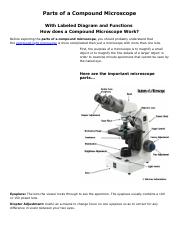
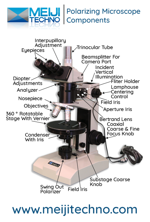
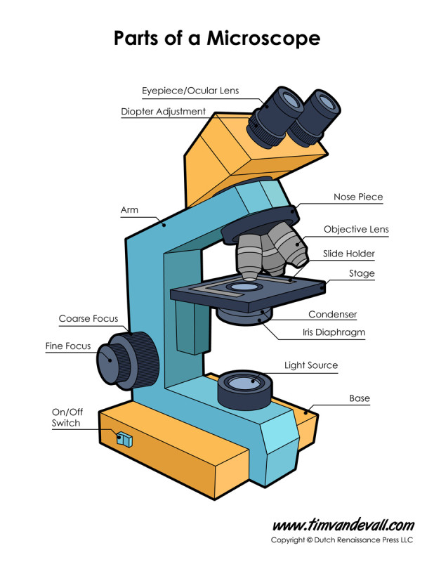
40 microscope diagram labeled parts
Cytology questions - Acaproso Name parts labeled A,B,C,D,E,F,G and H. Briefly explain how organelles A, C,D and H are involved in the synthesis and secretion of the inactive enzyme. Discussion (1) Discuss 3511. Category: (Short answers) Draw the structure of animal cell as seen under electron microscope i. Name a double membrane organelle found inplant cell only Veterinary Anatomy » AnatomyLearner >> The Place to Learn Veterinary ... The dog hip anatomy consists of bones, muscles, and joint. Some of the nerves and vessels supply to the hip region of a dog and pass it along. In this guide, I will tell (show) you the complete anatomical features of the dog's hip with the different labeled diagrams. Here, I will discuss the main … Read more
Spinal Cord Cross Section | New Health Advisor 1. White Matter The white matter has the nerve fibers that run up and down the length of the cord, they are called axons. This makes it possible for the different parts of the CNS communicate with each other. Every bundle of axons is a tract and it transmits specific information.

Microscope diagram labeled parts
Hypodermis (Subcutaneous Tissue): Anatomy and Function - Verywell Health Function. The functions of the hypodermis include: 4. Fat and energy storage: Fat cells (adipocytes) make up the fatty (adipose) tissue that stores energy for the body. The hypodermis also helps to create hormones such as estrogen and leptin. 3. Protecting the body: The fat in the hypodermis acts like padding or a shock absorber that protects ... Single-crystal X-ray Diffraction - Techniques What is Single-crystal X-ray Diffraction. Single-crystal X-ray Diffraction is a non-destructive analytical technique which provides detailed information about the internal lattice of crystalline substances, including unit cell dimensions, bond-lengths, bond-angles, and details of site-ordering. Directly related is single-crystal refinement ... BIOLOGY FORM ONE NOTES FREE - Educationnewshub.co.ke Anatomy: Study of structure of cells; Cytology: Study of cells; Biochemistry: ... MICROSCOPE. Microscope Parts & Function. Parts of the Microscope. 1. Eyepiece ... Diagrams Parts of a leaf. Lamina: This is the flat surface. It is green in colour and contain the photosynthetic tissue.
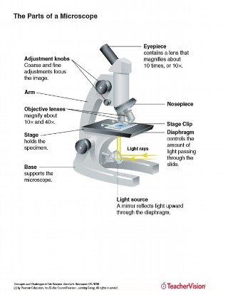
Microscope diagram labeled parts. Light Microscope (Theory) - Amrita Vishwa Vidyapeetham The modern compound microscope consists of two lens system, the objective and the ocular or eye piece. The first magnified image obtained with objective lens, is again magnified by the eye piece to give a virtual inverted image. The total magnification the product of the magnifications of two lens systems. Parts of a Microscope Telescope Meade Diagram Parts - swp.omeopatia.genova.it Search: Meade Telescope Parts Diagram. Applications Along with brands like Orion, Celestron and Sky-Watcher, Meade is one of the most trusted and popular names in the industry Very easy to use and much better than fiddling around using an allen key in the dark 4 QUICK-START GUIDE It is recommended that you attach the supplied tripod to the LX200GPS for observing SkyShed Roll Off Observatories ... Light compound microscope parts? [Expert Review] These parts include: eyepiece - also known as the ocular. this is the part used to look through the microscope. its found at the top of the. eyepiece tube - it's the eyepiece holder. it carries the eyepiece just above the objective lens. in some microscopes. objective lenses - these are the. Quiz Microscope Of Parts And Function [NH8XBD] Search: Parts Of Microscope And Function Quiz. Wednesday- 2/13: QOD: ID the numbered parts on the microscope on the SmartBoard These cells also accumulate at sites of infection, and the release of prostaglandins, serotonin and histamine help to increase blood flow to the area of damage, as part of the inflammatory response Explain the importance of each portion of the duct system and accessory ...
X-ray Powder Diffraction (XRD) - Techniques X-ray powder diffraction (XRD) is a rapid analytical technique primarily used for phase identification of a crystalline material and can provide information on unit cell dimensions. The analyzed material is finely ground, homogenized, and average bulk composition is determined. Fundamental Principles of X-ray Powder Diffraction (XRD) Simulations and Virtual Labs - Open Educational Resources - Library ... Provides the ability to explore the anatomy of a frog by using data from high resolution MRI imaging and mechanical sectioning, together with 3D surface and volume rendering software. ... Chemix is an online editor for drawing science lab diagrams and school experiment apparatus. The app provides easy sketching for both students and teachers ... scheme work biology - Free KCPE Past Papers Draw and label the light microscope; ... Drawing and labeling the light microscope . Light microscope; Diagram of light microscope; Comprehensive secondary Biology students Bk. 1 page 17; ... KLB teachers book 1 pages 23-25 . 10. 1-2. THE CELL. Parts of the light microscope and their functions . Calculation of magnification using light ... 30 Basic Parts of Boat, their Names, Functions & Diagram Different Parts of Boat Anatomy, Names & Diagram September 18, 2022 Swap Overview of Boat Anatomy Parts of Boat Diagram Parts of Boat Names Boat Parts Ballast Berth Bilge Bimini Bow Bulkhead Cabin Casting deck/platform Cleat Cockpit Console Deck Dinette Flybridge Galley Gunwale Hardtop Hatch Helm Hull Livewell Propeller Rigging Rudder Stern
Diagram Definition & Meaning - Merriam-Webster diagram: [verb] to represent by or put into the form of a diagram. X-Ray Generation Notes - University of Oklahoma X-Ray photons are electromagnetic radiation with wavelengths typically in the range 0.1 - 100 Å. X Rays used in diffraction experiments have wavelengths of 0.5 - 1.8 Å. X Rays can be produced by conventional generators, by synchrotrons, and by plasma sources. Electromagnetic radiation from nuclear reactions, called γ radiation, can also ... Microorganism - Wikipedia A microorganism, or microbe, is an organism of microscopic size, which may exist in its single-celled form or as a colony of cells.. The possible existence of unseen microbial life was suspected from ancient times, such as in Jain scriptures from sixth century BC India. The scientific study of microorganisms began with their observation under the microscope in the 1670s by Anton van Leeuwenhoek. 5 White Blood Cells Types and Their Functions - New Health Advisor Agranulocytes are free of visible grains under the microscope and include lymphocytes and monocytes. Together, they coordinate with one another to fight off things like cancer, cellular damage, and infectious diseases. Below, detailed information about each type will be discussed. 1. Neutrophils
Labeling the parts of a dissecting microscope Quiz
ECLIPSE Ti2 Series | Inverted Microscopes | Nikon Microscope Products ... The ECLIPSE Ti2 inverted microscope delivers an unparalleled 25mm field of view (FOV) that revolutionizes the way you see. With this incredible FOV, the Ti2 maximizes the sensor area of large-format CMOS cameras without making compromises, and significantly improves data throughput. The Ti2's exceptionally stable, drift-free platform is ...

Microscopes Microscope Parts Quiz on Friday!!
Famous Drawing Of Cross Section Of Leaf 2022 - Chic Way (I) Draw The Diagram Of Cross Section Of A Leaf And Label The Following Parts : Veins provides support to the leaf and veins consists of xylem and phloem. The tissues, in turn, are constructed of specialized cells, and the cells, of organelles. Short distance for carbon dioxide to. Biology Diagram For Cbse Class 10.

Produk Microscope | UD Berkah Abadi
Autoclave: Principle, Procedure, Types, Uses - Microbe Online Autoclave comprises three parts: a pressure chamber, a lid, and an electrical heater. Gravity Displacement type Autoclave Pressure chamber consists of - Large cylinder (vertical or horizontal) in which the materials to be sterilized are placed. It is made up of gunmetal or stainless steel and placed in a supporting iron case ay through

Microscope With Labels clip art | Microscope parts ...
Welcome to Boreal Science Printed from Boreal Science Website User: [Anonymous] Date: 09-19-2022 Time: 07:13
Microscope - Teaching resources
hypothalamus | Definition, Anatomy, & Function | Britannica hypothalamus, region of the brain lying below the thalamus and making up the floor of the third cerebral ventricle. The hypothalamus is an integral part of the brain. It is a small cone-shaped structure that projects downward from the brain, ending in the pituitary (infundibular) stalk, a tubular connection to the pituitary gland. The hypothalamus contains a control centre for many functions ...

Label the Microscope Diagram | Download Scientific Diagram
Gram Stain Technique (Theory) : Microbiology Virtual Lab I ... It is also a key procedure in the identification of bacteria based on staining characteristics, enabling the bacteria to be examined using a light microscope. The bacteria present in an unstained smear are invisible when viewed using a light microscope. Once stained, the morphology and arrangement of the bacteria may be observed as well.
MICROBIO 16 Parts of a Compound Microscope with Diagram and ...
Form 1-4 Biology Notes, Revision Questions and Answers ... Anatomy: Study of structure of cells; Cytology: Study of cells; Biochemistry: ... MICROSCOPE. Microscope Parts & Function. Parts of the Microscope. 1. Eyepiece ... Diagrams Parts of a leaf. Lamina: This is the flat surface. It is green in colour and contain the photosynthetic tissue.
Parts of a Microscope Quiz
Mr. Jones's Science Class Matter: Atoms and Properties - Open Response Question 3. Force and Motion - Open Response Question 3. Forms of Energy - Open Response Question 1. Forms of Energy - Open Response Question 2. Earth's Structure & Natural Processes - Open Response Question 1.
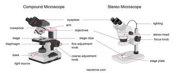
Parts of Stereo Microscope (Dissecting microscope) – labeled ...
Skin Layers: Structure, Function, Anatomy, and More - Verywell Health The dermis is split into two parts. 3 Papillary Dermis The papillary dermis is the thin, upper layer that contains capillaries (tiny blood vessels) that help regulate skin temperature and provide nutrients to the epidermis. This skin layer also contains: Meissner corpuscles , which are receptors that transmit sensations of delicate touch

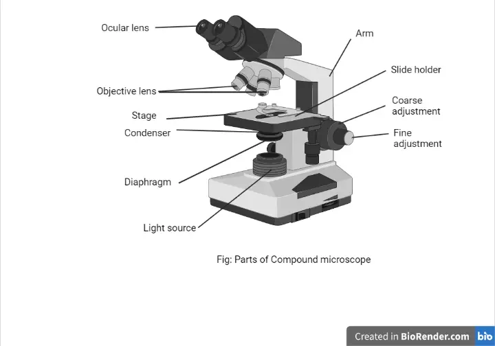
Parts of a microscope with functions and labeled diagram
Electron Microscope: Principle, Types, Applications Schematic diagram of Transmission Electron Microscope Like any ordinary microscope, the electron microscope also uses a light source, a combination of lenses to produce a magnified image, however, this vary slightly as compared to ordinary light microscope. Source of light: The source of light is replaced by a beam of very fast-moving electrons.

Compound Microscope Parts, Functions, and Labeled Diagram ...
Drosophila melanogaster - Wikipedia Drosophila melanogaster is a species of fly (the taxonomic order Diptera) in the family Drosophilidae.The species is often referred to as the fruit fly or lesser fruit fly, or less commonly the "vinegar fly" or "pomace fly". Starting with Charles W. Woodworth's 1901 proposal of the use of this species as a model organism, D. melanogaster continues to be widely used for biological research in ...

22 Parts Of a Microscope With Their Function And Labeled ...
BIOLOGY FORM ONE NOTES FREE - Educationnewshub.co.ke Anatomy: Study of structure of cells; Cytology: Study of cells; Biochemistry: ... MICROSCOPE. Microscope Parts & Function. Parts of the Microscope. 1. Eyepiece ... Diagrams Parts of a leaf. Lamina: This is the flat surface. It is green in colour and contain the photosynthetic tissue.

Parts of a Compound Microscope and Their Functions
Single-crystal X-ray Diffraction - Techniques What is Single-crystal X-ray Diffraction. Single-crystal X-ray Diffraction is a non-destructive analytical technique which provides detailed information about the internal lattice of crystalline substances, including unit cell dimensions, bond-lengths, bond-angles, and details of site-ordering. Directly related is single-crystal refinement ...

Parts of Microscope (Labeling) Diagram | Quizlet
Hypodermis (Subcutaneous Tissue): Anatomy and Function - Verywell Health Function. The functions of the hypodermis include: 4. Fat and energy storage: Fat cells (adipocytes) make up the fatty (adipose) tissue that stores energy for the body. The hypodermis also helps to create hormones such as estrogen and leptin. 3. Protecting the body: The fat in the hypodermis acts like padding or a shock absorber that protects ...

Dissecting Stereo Microscope Parts and Functions

File:Microscope diagram.png - Wikimedia Commons

Microscope, Microscope Parts, Labeled Diagram, and Functions
Parts of a Microscope with Their Functions – Microbe Online

microscope | Types, Parts, History, Diagram, & Facts | Britannica

Simple Microscope - Parts, Functions, Diagram and Labelling ...

Parts of the Microscope worksheet
Microscope Terminology

Labeled Microscope Diagram | Microscope parts, Science fair ...

Microscope Labeled Parts - ClipArt Best
Compound Microscope Parts, Functions, and Labeled Diagram ...
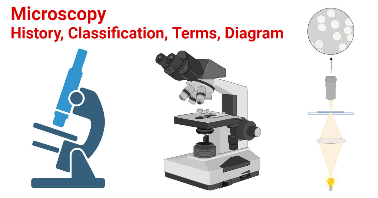
Microscopy- History, Classification, Terms, Diagram

Parts of the Microscope with Labeling (also Free Printouts ...

ABOUT MICROSCOPES | Scienceart

Microscope Parts and Functions

Tsetse biology, systematics and distribution, techniques

Parts of a microscope with functions and labeled diagram
Labelled Diagram of Microscope Parts
Microscope Diagram Labeled, Unlabeled and Blank | Parts of a ...
The Parts of a Microscope (Labeled) Printable Printable (6th ...

Compound Microscope Parts

Simple Microscope Definition, Magnification, Parts And Uses
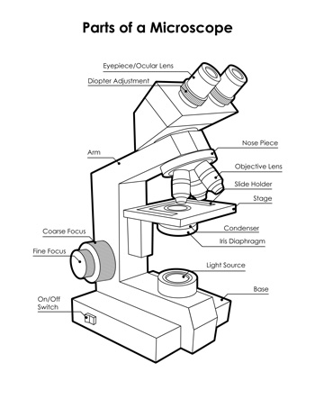
Labeled Microscope Diagram - Tim's Printables

Labelling a Microscope Diagram | Quizlet
Name Date Sci STANDARD MICROSCOPE DIAGRAM Label only the ...

Lasec Education | Key parts of a compound microscope and how ...
Post a Comment for "40 microscope diagram labeled parts"