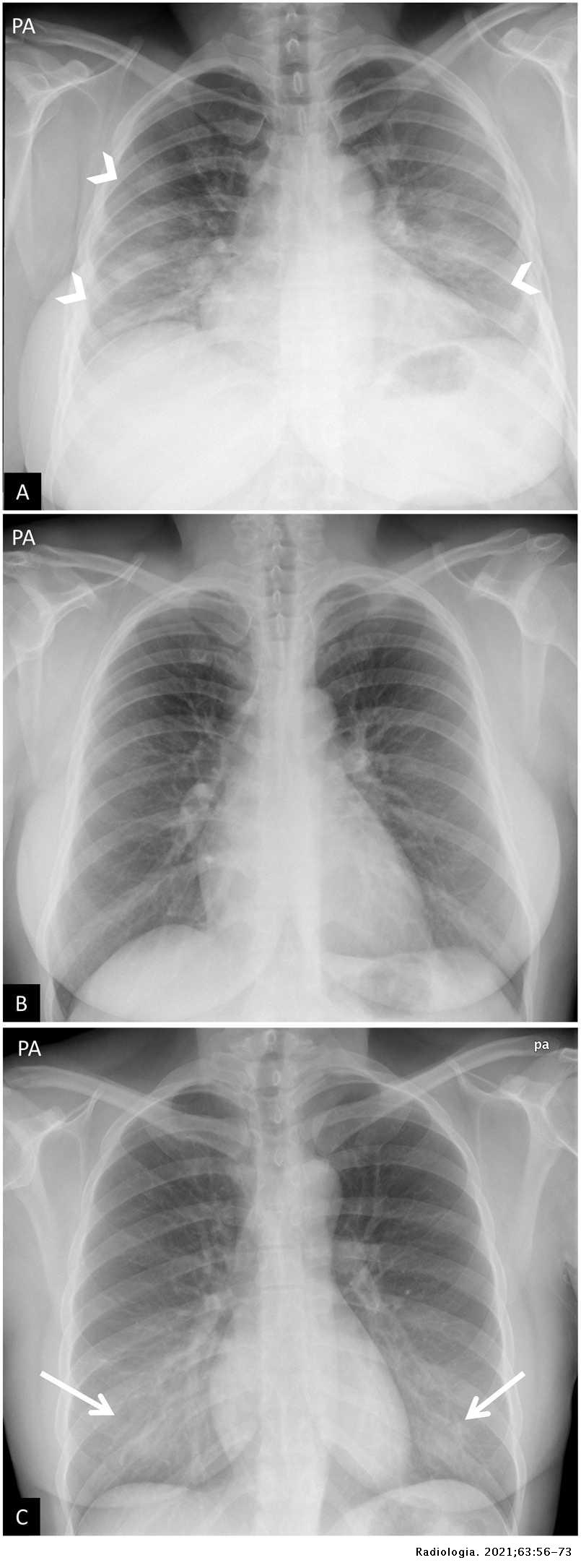
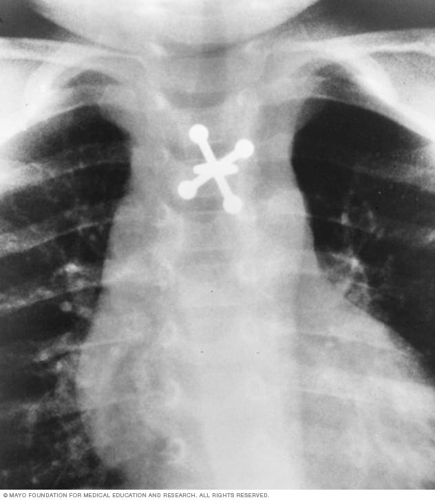
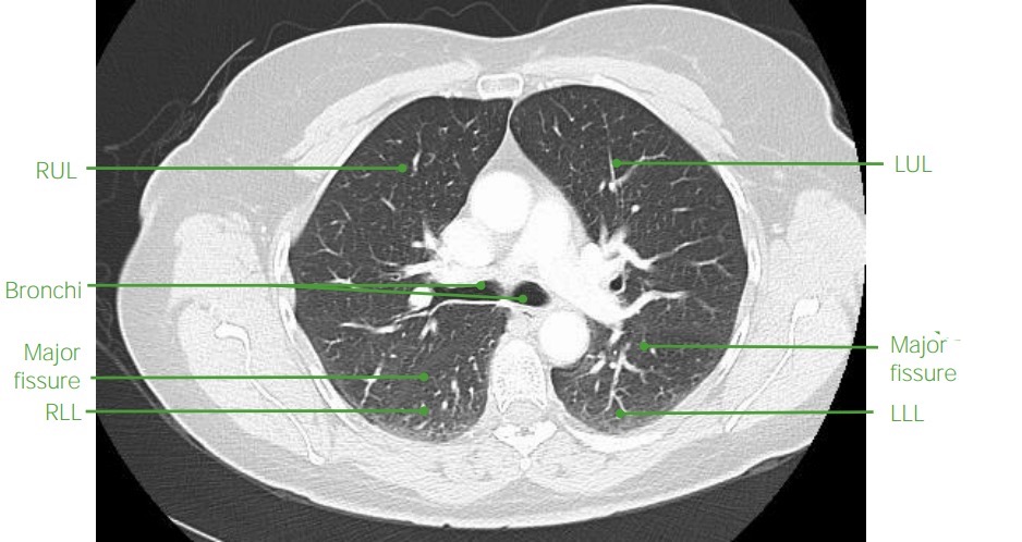
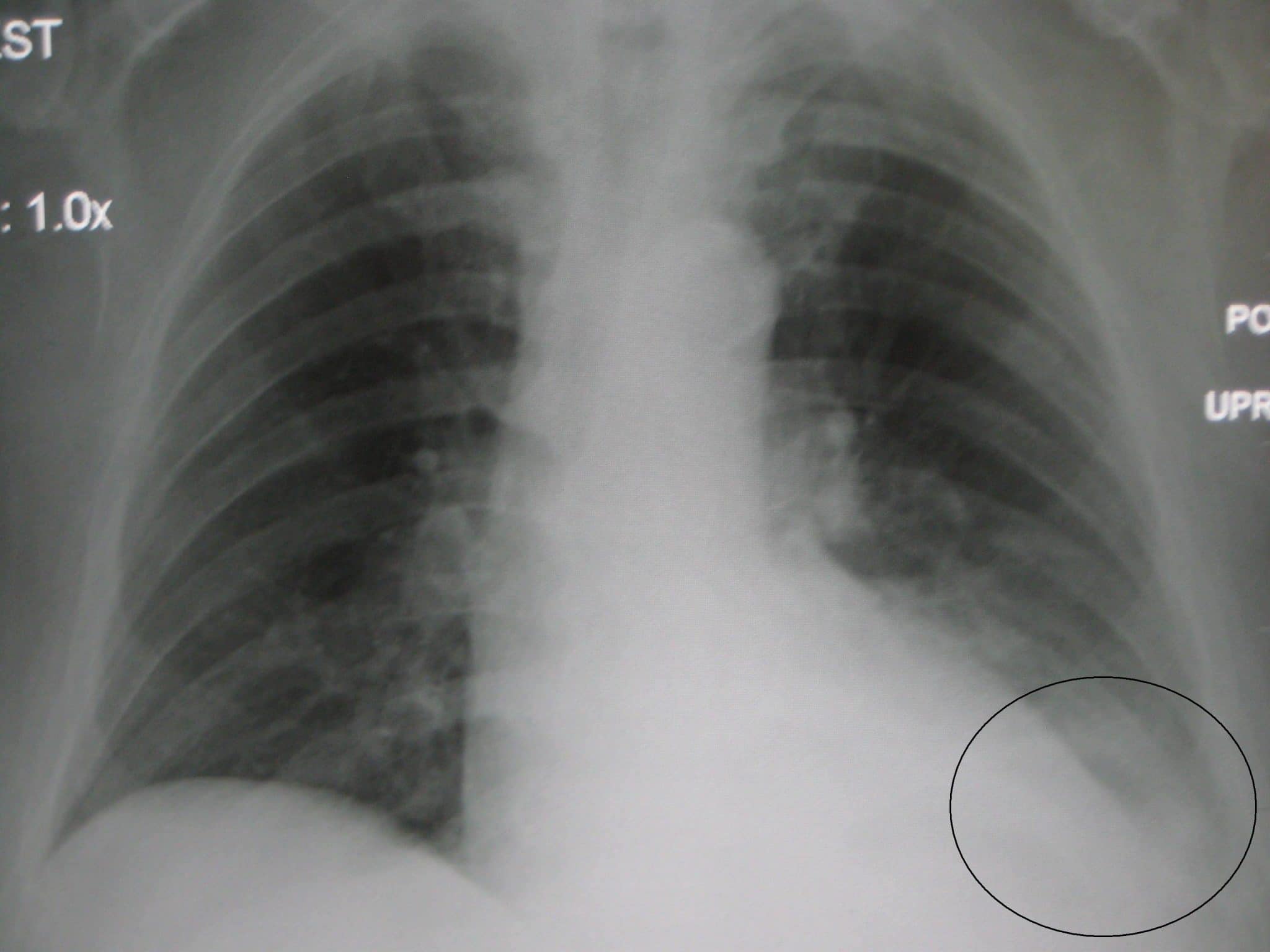
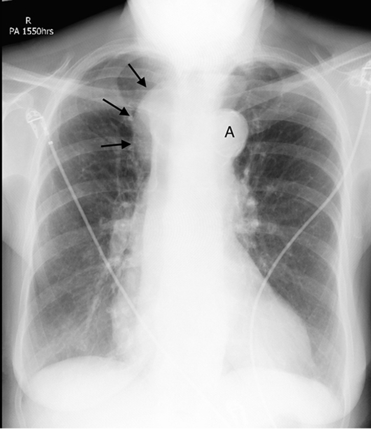
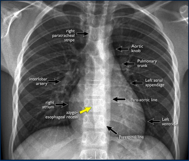
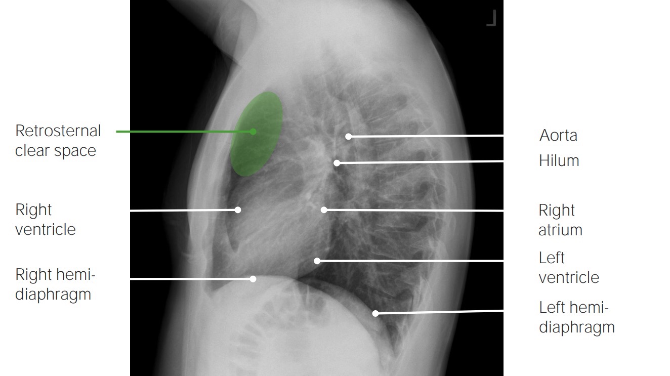
44 label the structures in the chest x-ray using the hints provided.
The Ultimate 12-Lead ECG Placement Guide (With Illustrations) To clarify, leads will equal: V4=V7, V5=V8, and V6=V9. Lastly, a right sided 12-lead ECG placement allows you to detect a right sided infarct. At a minimum, lead V4 should be placed on the 5th intercostal, mid-clavicular (exact opposite of the regular left side placement) if an inferior infarct was originally seen in leads II, III, and AVF. High-throughput phenotyping and genetic linkage of cortical bone ... Once the images are collected, the analysis is done using an in house post-processing pipeline (Fig. (Fig.1b,c) 1 b,c) to segment, label, and quantitatively characterize the structures. With new detector technology already enabling scan times below a second and continual improvements in the efficiency and performance of the entire pipeline, the ...
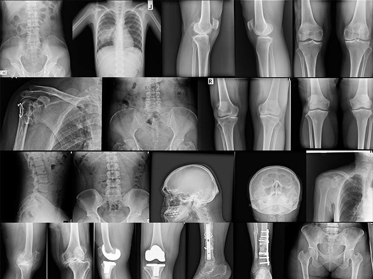
Principles of Bone X-Ray Diagnosis - 2nd Edition - Elsevier A chapter on widespread and regional reduction in bone density has been completely rewritten. This volume provides radiographs to illustrate every condition described. These radiographs were selected to illustrate the principles of bone x-ray diagnosis rather than to make a complete catalogue of all the conditions referred.
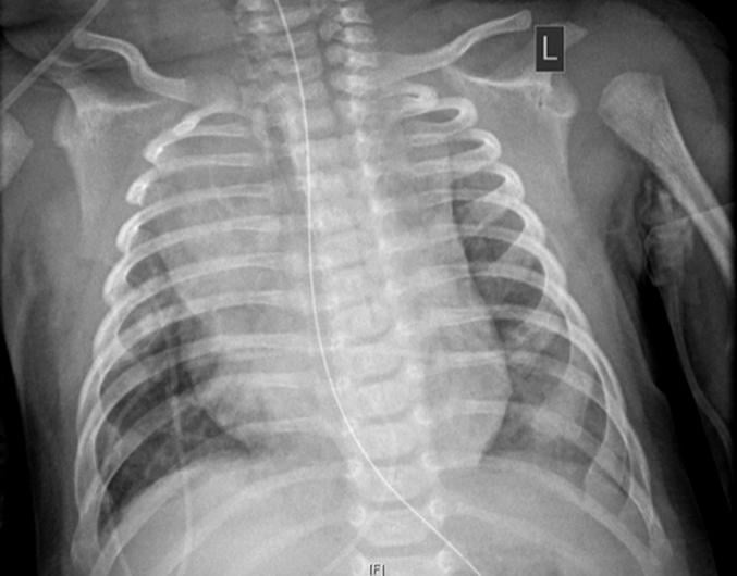
Label the structures in the chest x-ray using the hints provided.
Anatomical Directional Terms and Body Planes - ThoughtCo Some examples include the anterior and posterior pituitary, superior and inferior venae cavae, the median cerebral artery, and the axial skeleton. Affixes (word parts that are attached to base words) are also useful in describing the position of anatomical structures. These prefixes and suffixes give us hints about the locations of body structures. Solved Thorax X-ray -6 Label the structures in the chest | Chegg.com Best Answer 1) AORTIC KNOB 2) SUPERIOR VENA CAVA 3) RIGHT B … View the full answer Transcribed image text: Thorax X-ray -6 Label the structures in the chest X-ray using the hints provided Superior vena cava Aortic knob rences Apex of heart Right border of heart Loft border of heart Aorticoob Apex of heart Name the structure Reset Zoom ReaderUi ReaderUi
Label the structures in the chest x-ray using the hints provided.. (Solved) - Label the blood vessels using the hints provided. Posterior ... The blood vessels labelled ... [Solved] Drag the label to the appropriate box | Course Hero Step-by-step explanation Image transcriptions oxygenated Deoxygenated mixed area 1. Vessels depicted 1. Vessels depicted us in sued 1. Pulmonary Cappellaites 2. Left ventricle 2. Jugular vein 2. Systemic capillaries 3. Coronary arteries 3. Coronary sinus 4. Decending 4 Right Atrum 5. Carolid 5. Pulmonary arteries artery 6. Overview of the proposed automated chest X-ray reporting ... - ResearchGate Chest radiographs are one of the most common diagnostic modalities in clinical routine. It can be done cheaply, requires minimal equipment, and the image can be diagnosed by every radiologists.... Lab 9 Digestive and resp Flashcards | Quizlet Lab 9 Digestive and resp Term 1 / 156 Label the oral cavity and pharynx using the hints if provided. nasal septum nasopharynx tongue oropharynx epiglottis laryngopharynx esophagus Click the card to flip 👆 Definition 1 / 156 ... Click the card to flip 👆 Flashcards Learn Test Match Created by saruhhh1 Terms in this set (156)
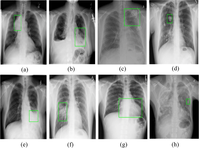
PDF Transfer Learning from CheXNet to COVID-19 The ChestX-ray14 contributes to our model as the dataset which our pre-trained layers were trained upon for CheXnet. The dataset was released by Wang et. al (2017) and contains 112,120 frontal- view X-ray images of 30,805 unique patients. Early diagnosis of lung cancer: which is the optimal choice? Chest x-ray. It is not recommended to use chest x-ray to detect lung cancer at present, owing to the difficulty of displaying intrapulmonary lesions and small lesions. Furthermore, whether combined with sputum cytology examination or not, chest x-ray screening cannot reduce lung cancer disease-specific mortality [5, 6]. Multi-modal trained artificial intelligence solution to triage chest X ... (1) dI dx = - μ ( x) I ( x) From this we can derive the beam intensity function over distance to be an exponential decay as the following: (2) I = I 0 e - ∫ μ ( x) dx In medical X-ray images the values saved to file are proportional to the expression seen in 3, which is actually a summation of the attenuation values along the beam. Lab 7 Blood & vessels Flashcards | Quizlet wall of inferior vena cava Place the following pictures of white blood cells (stained purple in the slides) into the appropriate category. highlighted structure- tunica externa of muscular artery highlighted structure- tunica media of muscular artery highlighted structure- tunica intima of muscular artery highlighted structure - arteriole
Body Systems - Definition and List of Body Systems - Toppr-guides It comprises of skin, hair, nails, sweat, and other glands which secrete substances onto the skin. 9. Lymphatic System or Immune System This system fights infection and includes lymphatic vessels which permeate the body. Every living thing needs to be able to fight infection. Clerking 101 | Symbols, Signs and Shorthand | Geeky Medics At this point, you should already be holding a pen with black ink and you should have ensured the continuation sheet has at least three key patient identifiers at the top. The next documentation steps include: 1. Adding the date and time (in 24-hour format) of your entry. 2. Writing your name and role as an underlined heading. 3. Select All That Apply NCLEX Practice Questions (100 Items ... - Nurseslabs Select All That Apply NCLEX Practice Questions and Tips (100 Items) Updated on May 25, 2022. By Gil Wayne, BSN, R.N. ADVERTISEMENTS. Practice answering select all that apply (SATA) questions for your NCLEX! Included in this free nursing test bank are 100 questions that are all multiple-response types covering different topics in nursing. Frequently Asked Questions - Contact & Support | LANDAUER Manage your account on myLDR your online account management tool for your dosimetry program. You can use myLDR to easily retrieve invoices and reports, make account changes, pay online and much more with this helpful tool. Sign up for an account at myLDR or contact our Client Experience Center at 800-323-8830.
Upper GI | Esophagram | Barium Swallow - Radiologyinfo.org Upper gastrointestinal tract radiography, also called an upper GI, is an x-ray examination of the esophagus, stomach and first part of the small intestine (also known as the duodenum). Images are produced using a special form of x-ray called fluoroscopy and an orally ingested contrast material such as barium.
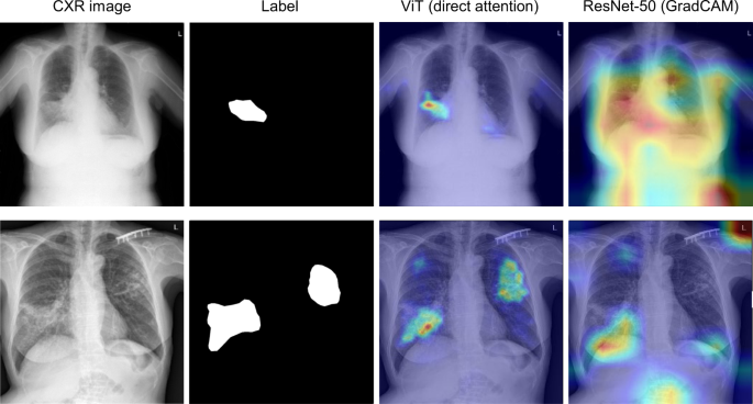
A Two-Step Radiologist-Like Approach for Covid-19 ... - SpringerLink We propose a two-step diagnostic approach for the detection of Covid-19 infection from Chest X-Ray images. Our approach is designed to mimic the diagnosis process of human radiologists: it detects objective radiological findings in the lungs, which are then employed for making a final Covid-19 diagnosis.
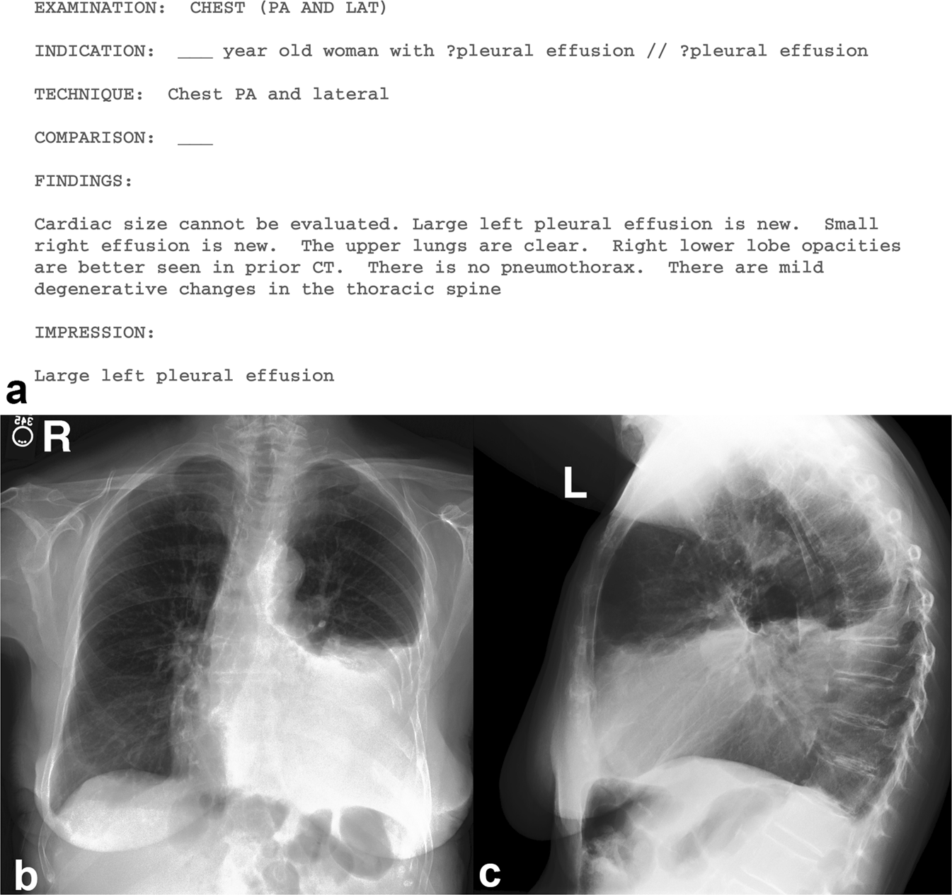
Frontal Radiograph | SpringerLink Understanding the basic components of a frontal view chest radiograph and its limitations is the cornerstone for mastering its literacy in order to successfully overcome those instances when the radiographs can challenge your confidence to diagnose. Keywords Frontal Heart borders Lung lobes Pleura AP PA Hilum Download chapter PDF Further Reading
The Lower Limb - TeachMeAnatomy The medical information on this site is provided as an information resource only, and is not to be used or relied on for any diagnostic or treatment purposes. This information is intended for medical education, and does not create any doctor-patient relationship, and should not be used as a substitute for professional diagnosis and treatment.
Module 3: Clinical Assessment, Diagnosis, and Treatment Module 3 covers the issues of clinical assessment, diagnosis, and treatment. We will define assessment and then describe key issues such as reliability, validity, standardization, and specific methods that are used. In terms of clinical diagnosis, we will discuss the two main classification systems used around the world - the DSM-5-TR and ICD-11.
Cardiovascular system general functions match each - Course Hero Hemostasis Drag each label to the appropriate position to indicate which step of hemostasis it describes. ... Label the structures in the chest X-ray using the hints provided. Right border of heart Apex of heart Superior vena cava Left border of heart ... statement of scope statement of authority Internet access policy acceptable use. document. 11.
PDF Utilizing Knowledge Distillation in Deep Learning for Classification of ... The overlapping of tissue structures in the X-ray images or the low contrast resolutions with which they need to distinguish the lesion and surrounding tissues greatly increase the complexity of...
5 Lung Cancer Nursing Care Plans - Nurseslabs Lung cancer is the most common cause of cancer death in men and women. Lung cancer is the carcinoma of the lungs characterized by uncontrolled growth of tissues of the lung. It usually develops within the wall or epithelium of the bronchial tree. Its most common types are epidermoid (squamous cell) carcinoma, small cell (oat cell) carcinoma, adenocarcinoma, and large cell (anaplastic) carcinoma.
Solved Thorax X-ray 79 Label the structures in the chest | Chegg.com Thorax X-ray 79 Label the structures in the chest x-ray using the hints provided points Aortic knob doc Apex of heart mos Superior vena CAVA Right bordet of heart Lott border of heart
Label the heart — Science Learning Hub In this interactive, you can label parts of the human heart. Drag and drop the text labels onto the boxes next to the diagram. Selecting or hovering over a box will highlight each area in the diagram. pulmonary vein semilunar valve right ventricle right atrium vena cava left atrium pulmonary artery aorta left ventricle Download Exercise Tweet
Step-by-step Guide to Building Your Own Neural Network From Scratch This structure is called a neuron. Schematic of a neuron. The function can be anything: a linear function or a sigmoid function. Of course, a single neuron has no advantage over a traditional machine learning algorithm. Therefore, a neural network combines multiples neurons. Think of neurons as the building blocks of a neural network.
ReaderUi ReaderUi
Solved Thorax X-ray -6 Label the structures in the chest | Chegg.com Best Answer 1) AORTIC KNOB 2) SUPERIOR VENA CAVA 3) RIGHT B … View the full answer Transcribed image text: Thorax X-ray -6 Label the structures in the chest X-ray using the hints provided Superior vena cava Aortic knob rences Apex of heart Right border of heart Loft border of heart Aorticoob Apex of heart Name the structure Reset Zoom
Anatomical Directional Terms and Body Planes - ThoughtCo Some examples include the anterior and posterior pituitary, superior and inferior venae cavae, the median cerebral artery, and the axial skeleton. Affixes (word parts that are attached to base words) are also useful in describing the position of anatomical structures. These prefixes and suffixes give us hints about the locations of body structures.
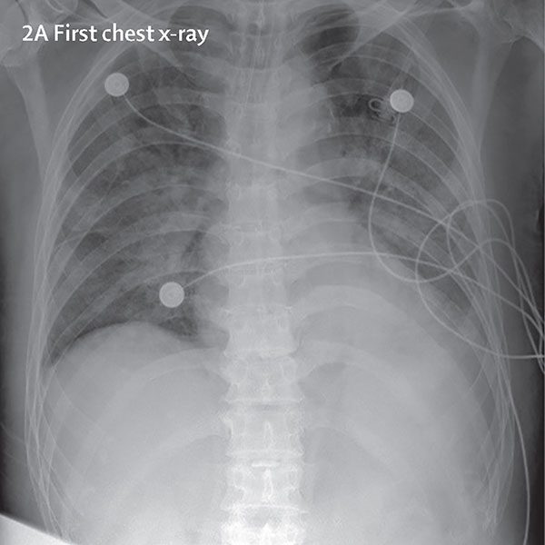





![20+ Best Image Datasets for Computer Vision [2022]](https://assets-global.website-files.com/5d7b77b063a9066d83e1209c/627d1222c7a13f26372caa1c_61252b50fc104417c2b9c8a4_computer-vision-datasets-hero.png)












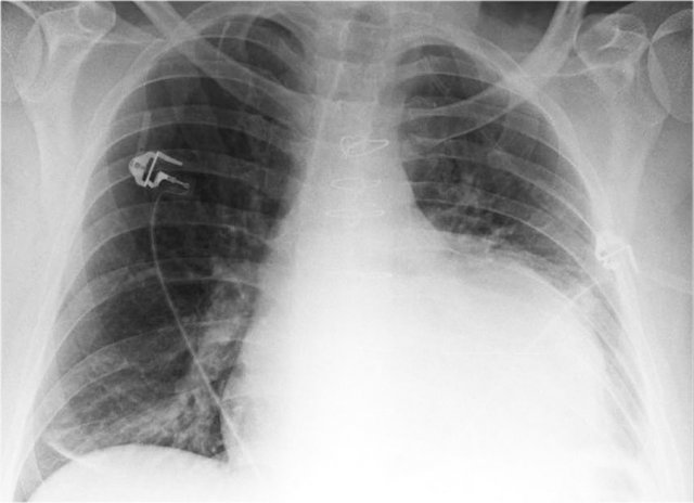
Post a Comment for "44 label the structures in the chest x-ray using the hints provided."